SciFig

Free Bacteria Diagram Generator

Create a labeled bacterial cell diagram, customize its structures and names, make a blank worksheet, and export SVG or PNG for your work.

Describe what you need in the box above.

Start with up to 150 free credits|No credit card required

Get up to 350 free credits on day one when you join through an invite.

3 figure engines · 500+ templatesEvery label editable · 60+ color schemesEditable PPTX · Layered SVG · 8K PNG / JPG

Build a bacterial cell diagram

Choose the cell shape, select its structures and edit the labels. Download your diagram or save the settings for later.

Cell shape
Structures and labels

Choose the structures to display and edit their names. Capsule, pili and flagellum are optional. Keep at least one structure selected.

Free Bacteria Diagram Generator— templates & examples

Updated

Bacterial cell diagrams

Free Bacteria Diagram Generator

Create a clear bacterial cell diagram with editable structures and labels.

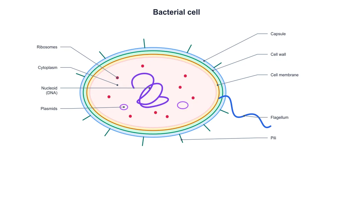
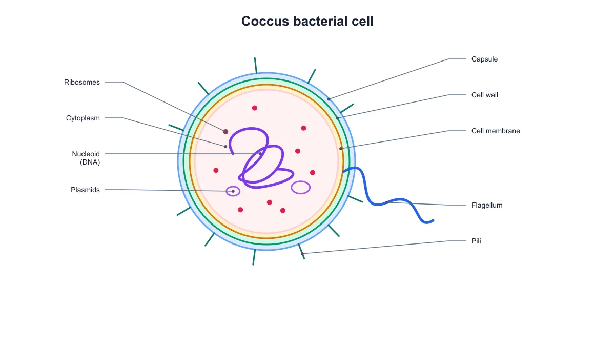
SciFig’s bacteria diagram generator helps you create a bacterial cell diagram with editable structure labels. Choose a rod-shaped cell or round coccus, select the parts to show and switch between a labeled reference and a blank worksheet. The drawing separates the cell envelope, nucleoid, plasmids and other selected structures. Export SVG for vector editing or PNG for a lesson, report or presentation, and save the settings to reopen the same figure later.

Labeled bacterial cell diagram with the capsule, cell wall, membrane, nucleoid, plasmids, ribosomes, pili and flagellum
  • Choose rod-shaped or round cells
  • Edit labels and optional structures
  • Make labeled or blank diagrams
  • Export SVG, PNG and saved settings

Bacteria Diagram Generator Features

Build a focused bacterial cell diagram and keep the figure with the settings that created it.

Start with a bacterial cell, not a blank canvas

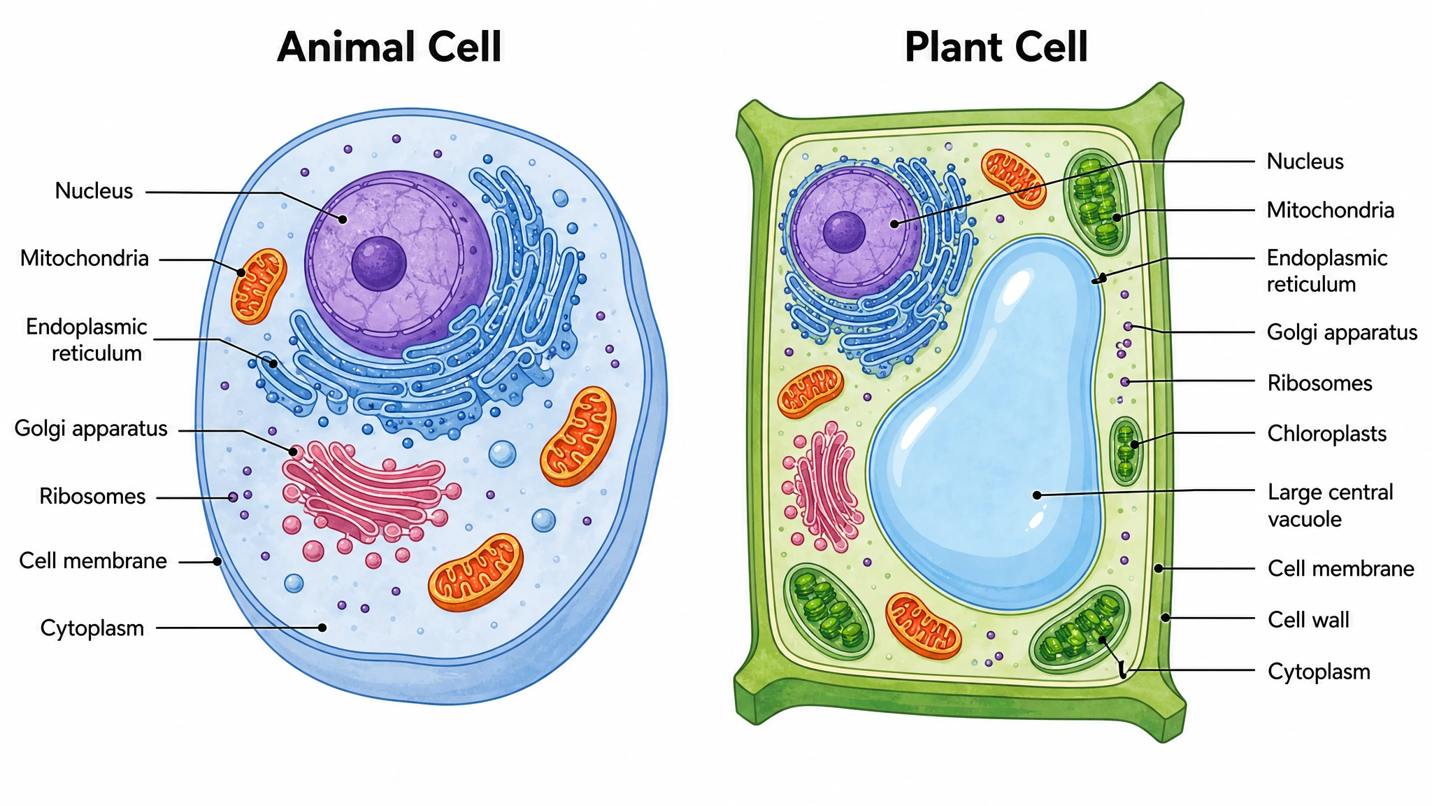
SciFig’s bacteria diagram generator starts with a labeled prokaryotic cell. Choose a rod-shaped cell or a round coccus, then select the structures your lesson or figure needs. The nucleoid holds the chromosomal DNA in the drawing; it is separate from the small plasmid rings. There is no membrane-bound nucleus to confuse this bacterial cell with an animal or plant cell.

  • Choose rod or coccus
  • Keep chromosomal DNA and plasmids distinct
  • Select the structures to display
Try this in SciFig

Edit each structure label and its visibility

Use the structure controls to show or hide the capsule, cell wall, cell membrane, cytoplasm, nucleoid, plasmids, ribosomes, pili and flagellum. SciFig keeps the selected structures and their leader lines together when you make a change. Customize the names for a lecture or handout, such as changing “Nucleoid (DNA)” to “Chromosomal DNA,” without redrawing the cell or repositioning every annotation.

  • Edit nine structure names
  • Keep leader lines with their parts
  • Choose optional appendages
Try this in SciFig

Make a blank labeling worksheet

Turn off labels to keep the bacterial cell drawing and its leader lines without the answers. SciFig uses the same structure selection for the labeled and blank versions, so a teaching slide and a student worksheet can share one diagram. Select fewer structures for an introductory exercise, then return to the fuller diagram when the lesson moves to plasmids, pili or the cell envelope.

  • Switch labels on or off
  • Keep the same diagram geometry
  • Prepare focused labeling exercises
Try this in SciFig

Export your figure and keep its editable settings

Download the bacterial cell diagram as SVG for vector editing or PNG for a slide, worksheet or document. Save a JSON settings file to retain the selected shape, structure visibility, custom title and labels. Reopen that file to continue the same diagram later. SciFig AI Illustration (AI scientific illustration) also helps you create and refine other scientific illustrations from a description.

  • Download SVG or PNG
  • Save and reopen diagram settings
  • Continue with SciFig AI Illustration
Try this in SciFig

How to Make a Bacterial Cell Diagram

Choose the structures for your example, check the preview and export the figure you need.

  1. Choose a bacterial cell shape

    Start with the rod-shaped example or select coccus for a round cell. Give the diagram a title that matches the lesson, report or presentation. The selected shape updates the drawing immediately.

  2. Select the structures and edit their names

    Choose which structures to show and enter the labels you want to use. Capsule, pili and flagella are optional features rather than universal features of all bacteria. Keep the relevant structures for your example.

  3. Prepare a labeled figure or blank worksheet

    Leave labels on for an explanatory diagram, or turn them off for a student exercise. Check the live preview while changing the structure selection. The blank version retains the drawing and leader lines.

  4. Download and save the source settings

    Export SVG or PNG for your document. Save the JSON settings alongside the figure when you want to reopen and revise the same diagram. You can restore the title, labels, shape and visible structures together.

Bacterial Cell Diagrams for Teaching and Research

Make the diagram match the cell structures, level of detail and output your task calls for.

Labeled bacterial cell diagram with the capsule, cell wall, membrane, nucleoid, plasmids, ribosomes, pili and flagellum

Introduce bacterial cell anatomy

Explain the difference between a prokaryotic cell and a eukaryotic cell with an example that shows a nucleoid instead of a nucleus. Point to the membrane, cytoplasm and ribosomes while connecting each visible structure to its role. Export the labeled diagram for a lecture or study handout.

Explore this example
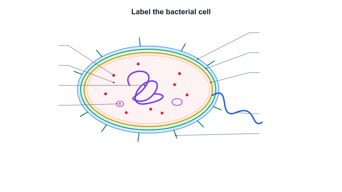
Blank bacterial cell diagram retaining its structural shapes and leader lines for a labeling worksheet

Make a labeling worksheet

Use the bacteria diagram generator to prepare the labeled reference and blank exercise from one set of settings. Hide the labels while keeping the leader lines so students can identify the parts. Save the settings when you want the same layout for a later answer sheet.

Explore this example
Round bacterial cell diagram showing the nucleoid and other selected prokaryotic structures

Compare bacterial shapes

Show a rod-shaped cell and a round coccus using the same selected structures and label wording. The paired diagrams help separate cell shape from the structural parts students are learning. Use the exported figures side by side in a slide or revision guide.

Explore this example

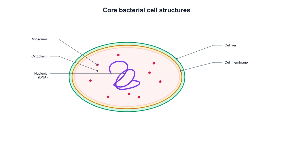
Bacterial cell diagram with the cell wall, membrane, cytoplasm, nucleoid and ribosomes

Focus a first microbiology lesson

Select the cell wall, membrane, cytoplasm, nucleoid and ribosomes for a less crowded introductory diagram. Introduce optional structures in a later version, using the same drawing controls. This gives a lesson a consistent visual vocabulary without starting again from an empty canvas.

Explore this example
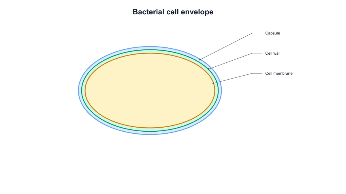
Bacterial cell envelope diagram with nested capsule, cell wall and cell membrane layers

Explain the cell envelope

Display the capsule, cell wall and cell membrane as separate nested layers. Use the custom labels to connect the figure to your teaching terminology, and keep the diagram focused on those outer structures. The exported SVG preserves separate drawing elements for further layout work.

Explore this example
Labeled bacterial cell diagram with the capsule, cell wall, membrane, nucleoid, plasmids, ribosomes, pili and flagellum

Prepare a scientific presentation

Create a clean bacterial cell figure for an introductory methods slide, teaching seminar or research overview. Edit the title and structure names, export the figure, and keep its source settings with the presentation. For more elaborate scientific scenes, continue in SciFig AI Illustration (AI scientific illustration).

Explore this example
How Researchers Use SciFig
I was preparing an introductory slide on prokaryotic cells and wanted the diagram to match the vocabulary in our course notes. The bacteria diagram generator let me select the structures and edit the names in one place. I exported the labeled figure for the lecture and kept a blank version for the student worksheet.
Maya Chen

Microbiology lecturer, University of Leeds

Free tools

A tool for every scientific figure

110 focused generators for the scientific figure you need — each opens in the right workspace with a matching example.

Bacteria Diagram Questions

1.

2.

3.

4.

5.

6.

Call to action background

Bacterial cell diagrams

Make your bacterial cell diagram

Create a bacterial cell diagram

Free to start · No credit card required · Built for researchers