Animal Cell Diagram Generator
Generate accurate animal cell diagrams — labeled or unlabeled — with nucleus, mitochondria, Golgi apparatus, endoplasmic reticulum, and key organelles for biology or research.
Figure prompt
Core Subject (e.g., Cas9 protein cutting DNA)
Action / Details (e.g., Double strand break, detailed molecular view)
Start with 100 free credits|No credit card required
Get up to 300 free credits on day one when you join through an invite.
Reviewed by SciFig TeamUpdated
Animal Cell Diagram Generator— templates & examples
Everything you need to generate your animal cell diagram
Labeled or unlabeled — your choice
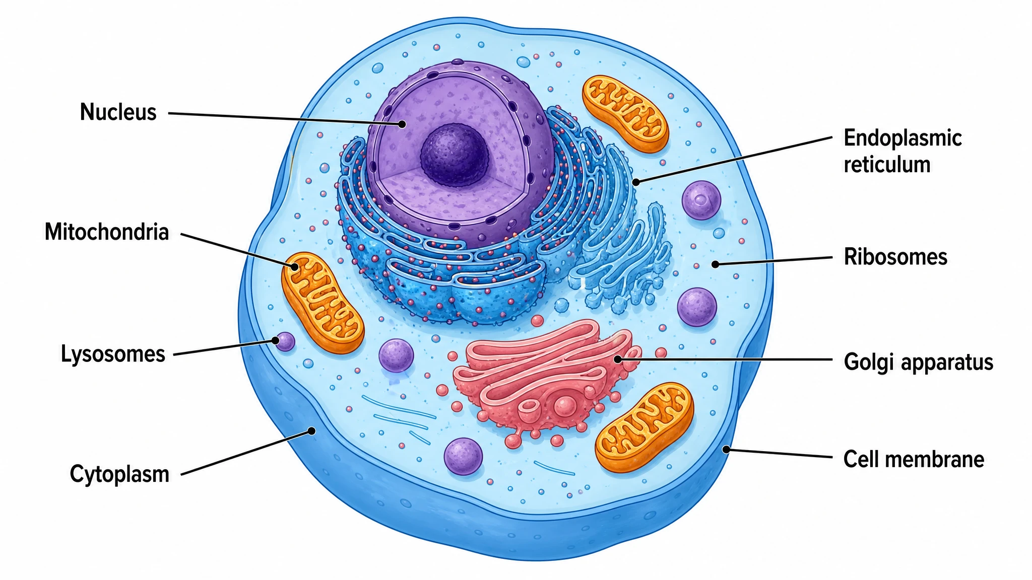
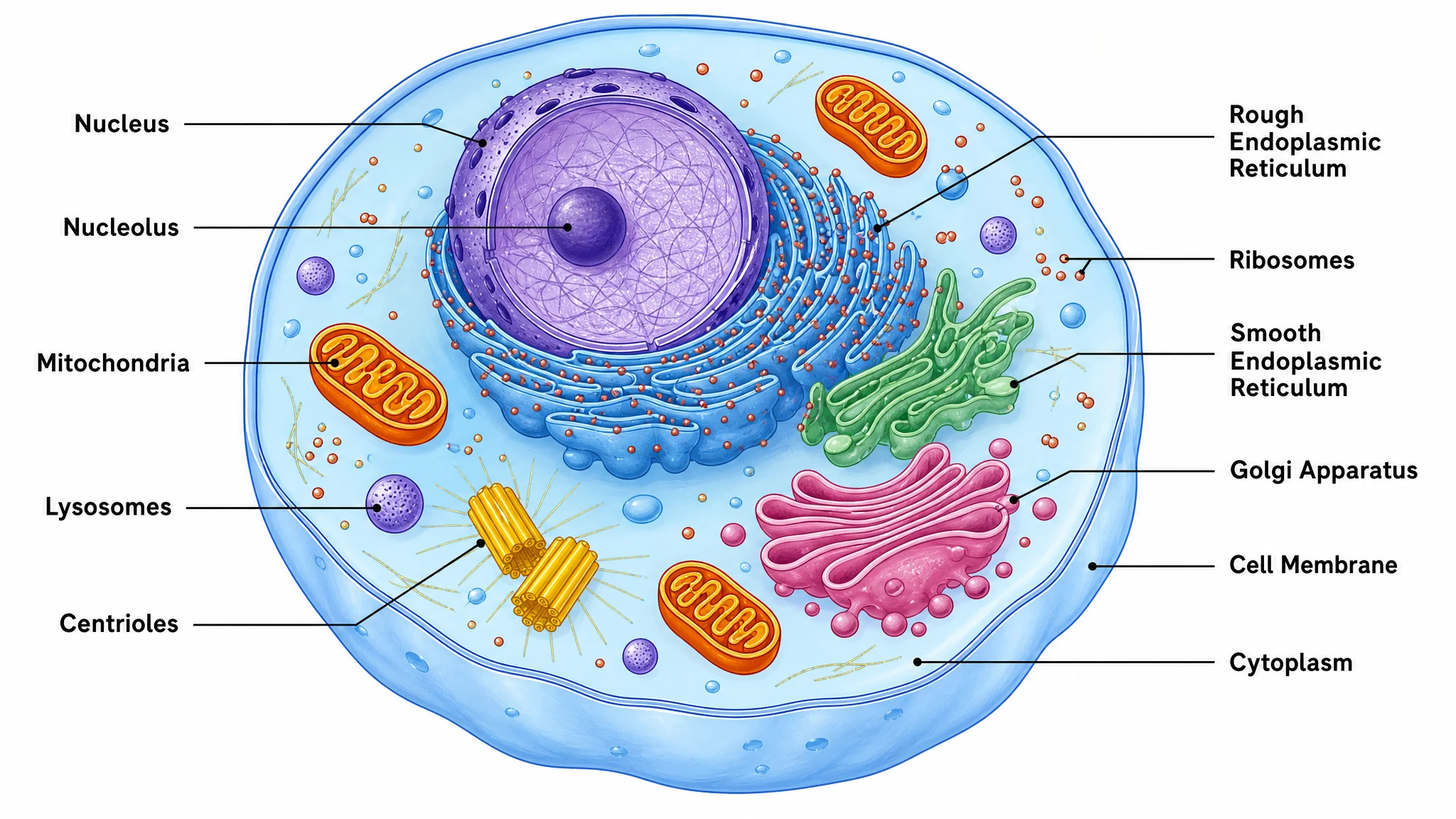
Whether you need a fully labeled animal cell diagram for a textbook figure or an unlabeled animal cell diagram for a student worksheet, SciFig produces both from the same description. Each organelle — nucleus, mitochondria, endoplasmic reticulum, Golgi apparatus, ribosomes, lysosomes, and cell membrane — is rendered accurately and consistently. Switch between labeled and blank versions without rebuilding anything.
Accurate biology, publication-ready style
SciFig's animal cell diagram generator is trained on biology illustration conventions, so every eukaryotic animal cell cross-section reflects real organelle shapes, positions, and proportions. Ask for an animal cell diagram labeled with leader lines and the callouts sit outside the cell body, annotating the organelles instead of covering them. The result is a clean, professionally styled figure suitable for peer-reviewed manuscripts, grant applications, lecture slides, and high-school or university course materials — no manual illustration skill required.
Instant cell diagram worksheets
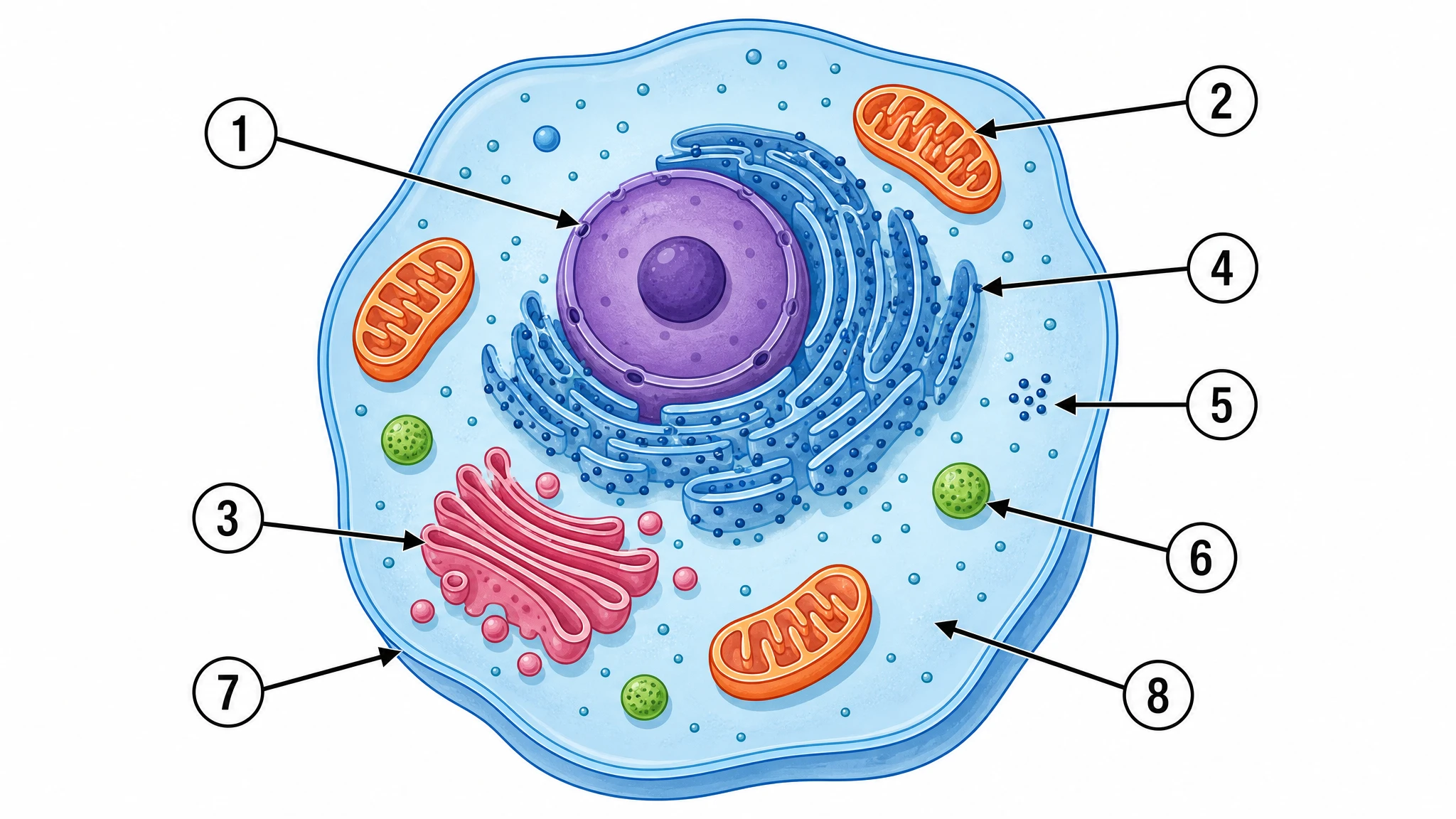
Create a complete cell diagram worksheet in under a minute: generate an unlabeled animal cell diagram with numbered callouts, then export the matching labeled answer key. Because both files come from the same render, an animal cell diagram unlabeled for the classroom matches its answer key organelle for organelle, with no drift between the worksheet and the key. Adjust the level of detail — a simple animal cell diagram for younger students or a richly detailed version for advanced biology — and download both as high-resolution images ready to print or embed in a slide deck.
What is an animal cell diagram?
An animal cell diagram is a labeled illustration of a eukaryotic animal cell in cross-section, showing organelles such as the nucleus, mitochondria, endoplasmic reticulum, Golgi apparatus, ribosomes, lysosomes, cell membrane, and cytoplasm. It is a core tool in biology education and research. SciFig's animal cell diagram generator produces a professionally drawn, fully editable figure in seconds — choose a labeled version for slides or an unlabeled blank for worksheets, then export.
Why an accurate animal cell diagram matters
- Visual diagrams accelerate comprehension of complex organelle functions compared to text descriptions alone
- Labeled animal cell diagrams are required figures in biology textbooks, lab reports, and peer-reviewed papers
- An unlabeled animal cell diagram — the animal cell blank diagram students fill in themselves — is one of the most commonly assigned biology worksheet formats worldwide
- Accurate organelle placement (nucleus, mitochondria, Golgi apparatus) prevents misconceptions that persist into higher education
- Publication-quality figures signal scientific rigor to journal reviewers and grant committees
- Editable diagrams let educators update organelle labels as curriculum standards evolve
Key organelles in an animal cell diagram
- Nucleus — membrane-bound control center containing DNA, nucleolus, and chromatin
- Mitochondria — double-membraned organelles that generate ATP through cellular respiration
- Endoplasmic reticulum — rough ER (protein synthesis) and smooth ER (lipid synthesis)
- Golgi apparatus — flattened membrane stacks that package, modify, and dispatch proteins
- Ribosomes — complexes on rough ER and in cytoplasm that translate mRNA into proteins
- Lysosomes — membrane-bound sacs of digestive enzymes that break down cellular waste
- Cell membrane — phospholipid bilayer that acts as a selective barrier around the cell
Where animal cell diagrams are used
- Middle school and high school biology courses covering eukaryotic cell structure
- University cell biology, molecular biology, and biochemistry lectures and exams
- Biology worksheet packets and standardized test preparation materials
- Peer-reviewed journal articles and review papers on cellular processes
- Grant proposals and research presentations requiring clear cell biology figures
- Online learning platforms, textbooks, and open educational resources
How to make an animal cell diagram
Describe your animal cell diagram
Tell SciFig what to draw in plain language — no design tools required.
Generate with SciFig
Get a clean, publication-ready figure that matches your description in seconds.
Edit & export
Vectorize it into editable SVG, relabel everything, and export for your paper, poster, or slides.
Animal Cell Diagram Generator — Frequently Asked Questions
Common questions about Animal Cell Diagram Generator.
More tools
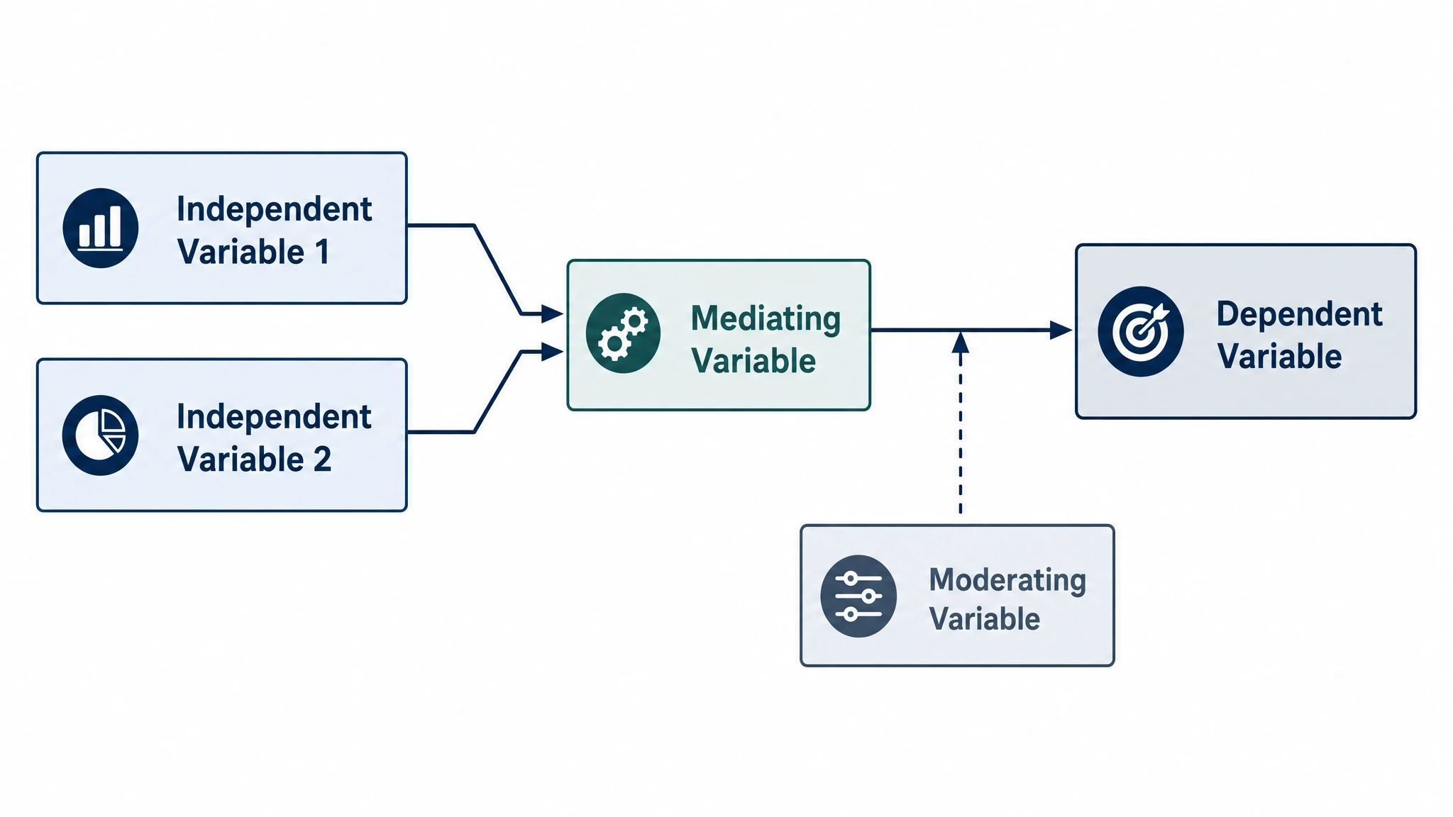
Conceptual Framework Generator
Create professional conceptual framework diagrams showing variable relationships, hypotheses, and theoretical models for your research.
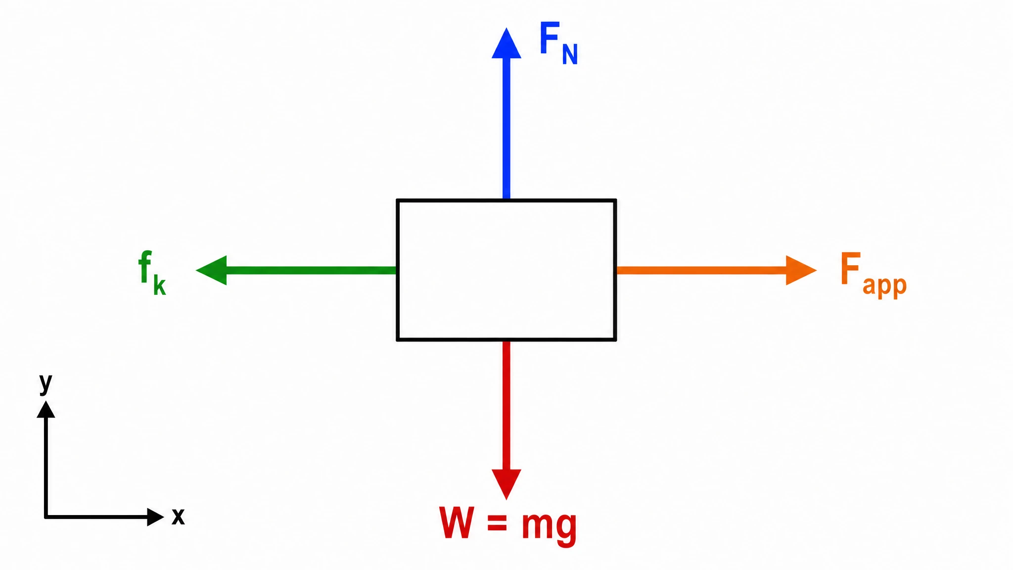
Free Body Diagram Generator
Create accurate, publication-ready free body diagrams with labeled force vectors for weight, normal force, friction, and tension in seconds.
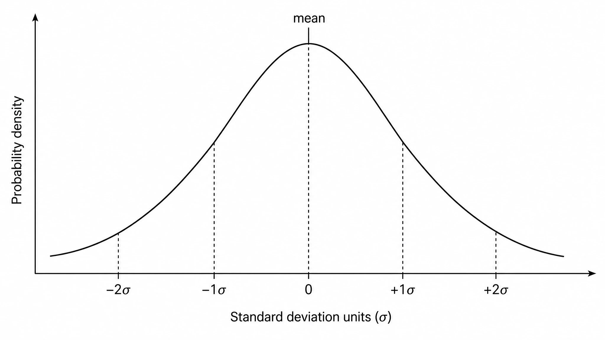
Bell Curve Generator
Generate a precise, fully labeled bell curve showing mean, standard deviations, and percentile regions — ready to export for your paper, thesis, or classroom.
Related links
Home
Turn text, sketches, references, PDFs, and photos into Scientific Figures
Text-to-Figure
Generate a figure from a plain-language description
Models
Default to GPT Image 2 for journal papers; switch to Nano Banana Pro for slides and posters; pick Nano Banana 2 for routine figure work
Tutorials
Seven short walkthroughs of the AI scientific figure generator
Blog
Tutorials, tool comparisons, and publication tips for researchers.
Inspiration
Explore publication-ready scientific figure examples, copy the prompts, and use them as starting points for your own work.

Ready to publish?
Make your own animal cell diagram in minutes.
Start for freeFree to start · No credit card required · Built for researchers