RSNA 2026 Poster & Presentation Guidelines and Template
RSNA 2026 poster and presentation guidelines: the Nov 29-Dec 3 dates, McCormick Place venue, the Aug 5 abstract deadline, and the radiology figures you need.
SciFig Team
SciFig editorial team
RSNA 2026 runs November 29 – December 3, 2026 at McCormick Place in Chicago, Illinois.
This is a practical guide to the official presentation rules for the Radiological Society of North America's 112th Scientific Assembly and Annual Meeting: the digital poster and education exhibit format, the abstract submission deadline and where accepted work is uploaded, and scientific paper (oral) slides.
It also covers the explanatory figures and graphical abstracts that make a radiology poster or slide worth stopping for, where accepted work is published, and the disclosure and AI questions still to be confirmed. Every date and specification below links to official RSNA materials.
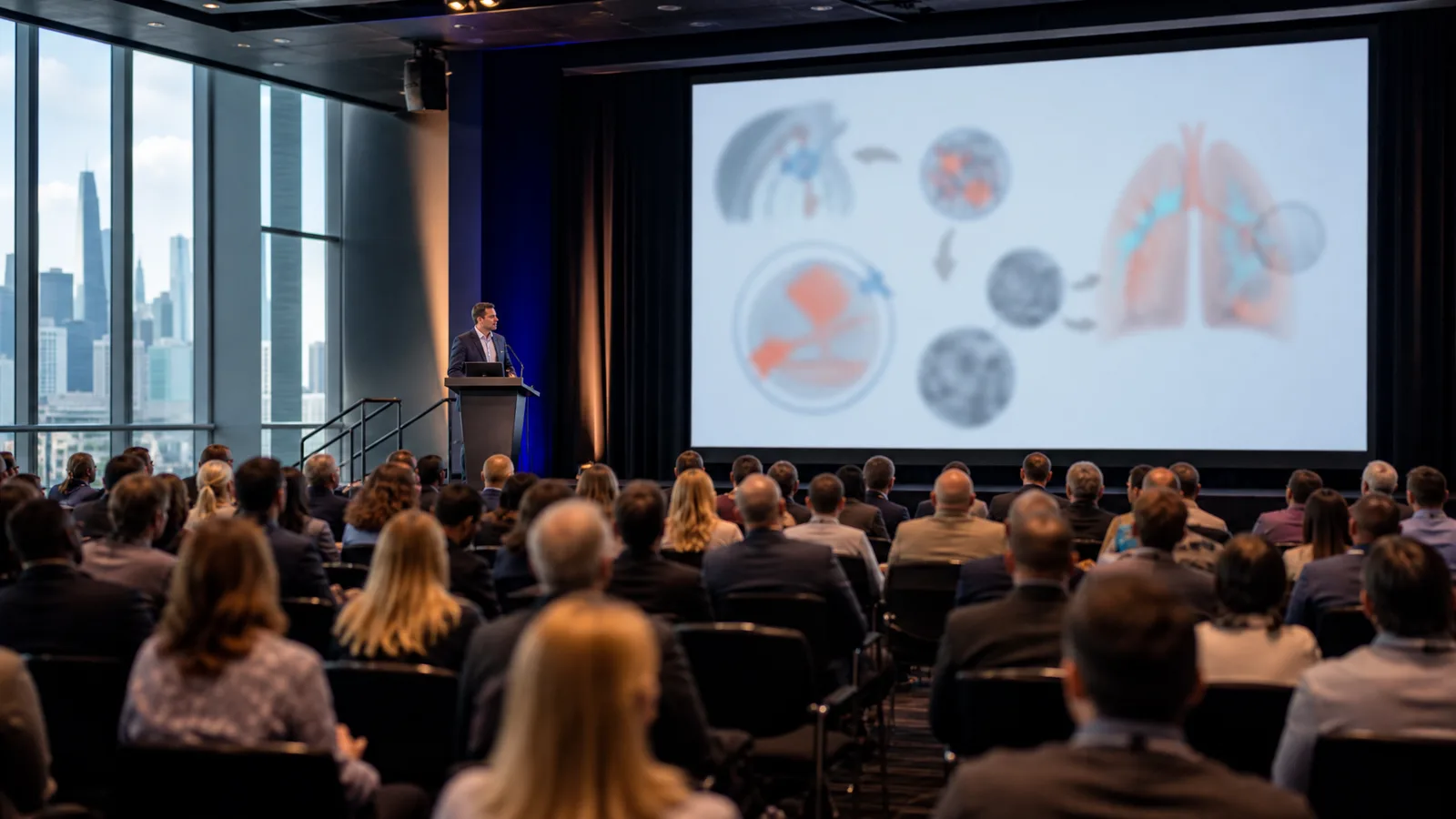
RSNA 2026 scientific session presentation: a presenter at the podium beside a large screen showing an imaging-study schematic, audience seated in a session hall (Figure generated with SciFig)
Transparency note: Illustrations in this article were generated with SciFig AI and reviewed by the author for scientific accuracy. Factual claims about dates and specifications link to official RSNA materials, and specifications RSNA has not yet published are flagged as such.
1. RSNA 2026 at a Glance: Dates, Venue, and Key Deadlines
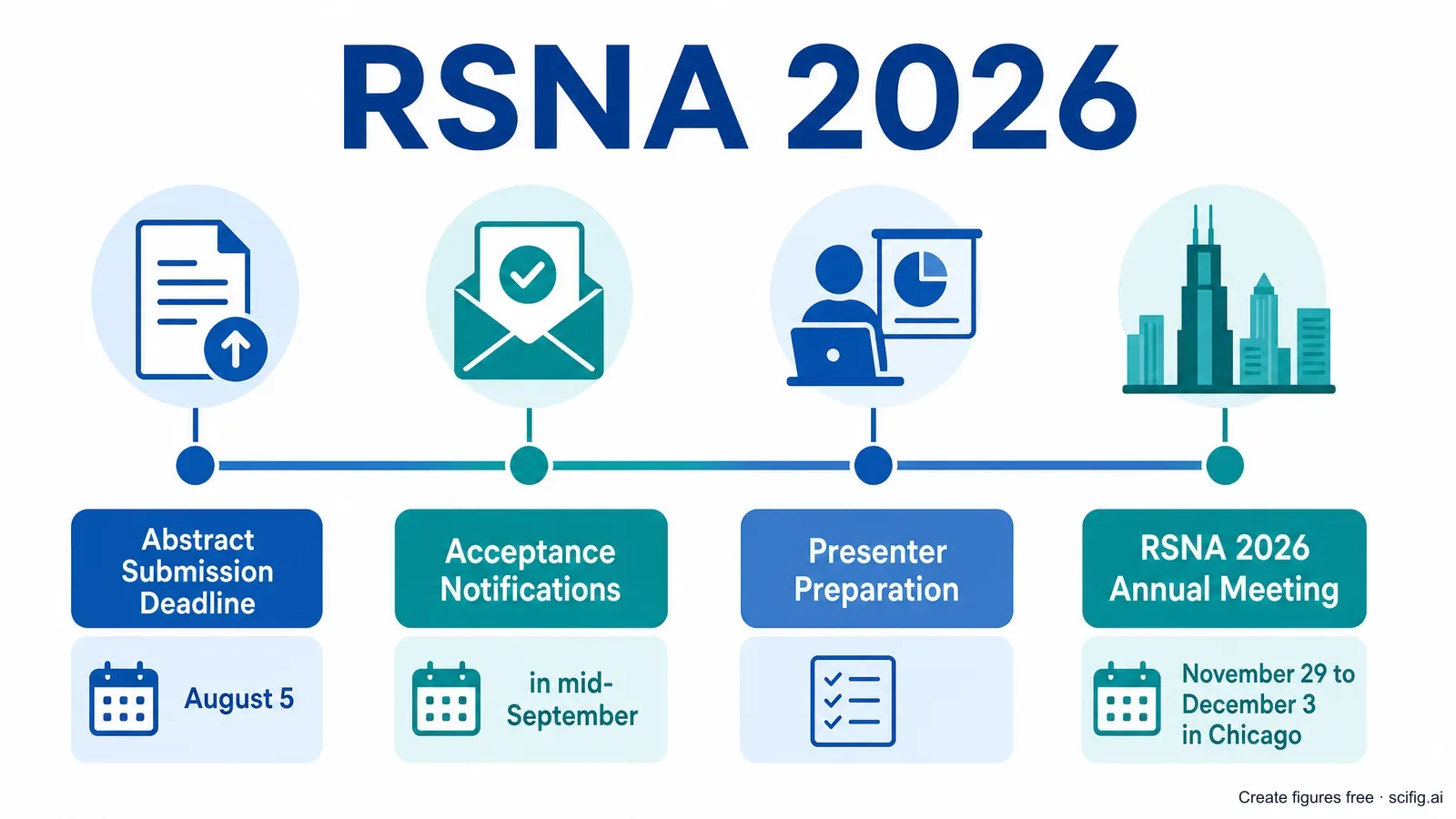
RSNA 2026 — the 112th Scientific Assembly and Annual Meeting of the Radiological Society of North America — runs November 29 – December 3, 2026 at McCormick Place in Chicago, Illinois (Central Time), under the theme "At the Center of Care." Abstracts are submitted in two categories, science and education, and the Cutting-Edge Research submission deadline is August 5, 2026, at 12 p.m. CT. Acceptance notifications are sent by email in mid-September 2026, and accepted presenters receive 50% off in-person registration — so it is worth mapping the full RSNA 2026 timeline early.
Here is the RSNA 2026 timeline that matters, drawn from the official RSNA 2026 annual meeting and RSNA abstract submission pages. For the session-by-session schedule, RSNA posts the program online closer to the meeting.
RSNA 2026 submission timeline: Cutting-Edge Research abstract deadline August 5, acceptance notifications mid-September, annual meeting November 29-December 3 (Figure generated with SciFig)
Date
Milestone
August 5, 2026 (12 p.m. CT)
Cutting-Edge Research abstract submission deadline
RSNA 2026 Annual Meeting, McCormick Place, Chicago
Accepted presenters receive 50% off in-person registration, per the official RSNA abstract submission page. Confirm your presentation and register once your notification arrives.
2. RSNA 2026 Poster Format and Guidelines: Digital, Scientific, and Education Exhibits
RSNA is a largely digital meeting, and its 2026 poster and exhibit format specifications — digital poster dimensions and file format, and education exhibit specs — are not published as of July 21, 2026; confirm them in the official RSNA presenter resources once acceptance notifications go out in mid-September. What is confirmed is the structure: abstracts are submitted in two categories, science and education, and each follows its own presentation track. Do not assume a printed-poster size here — RSNA is highly digitized, and the 2026 format (digital, printed, or hybrid) should be verified against official materials rather than borrowed from another society's rules.
The submission categories and awards come straight from the official RSNA abstract submission page; the detailed 2026 format specifications are typically released to accepted presenters after mid-September, in the RSNA event guidelines and presenter resources. A few points shape how you plan the sheet:
Two abstract categories — science and education — each with its own presentation format, so confirm which specs apply to your accepted work.
2026 dimensions and file format are not yet published — build your content and figures now, and set the final canvas size once official specs are released.
Design for screens — as a digital-first meeting, your work is likely to be read on a display, where crisp vector-style figures stay legible when a viewer zooms in.
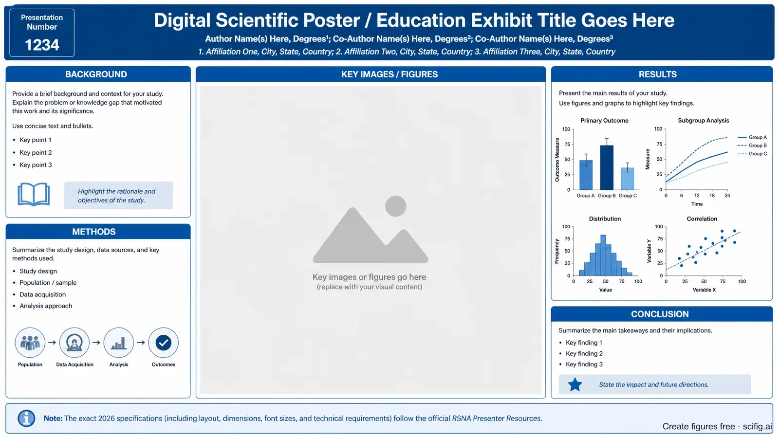
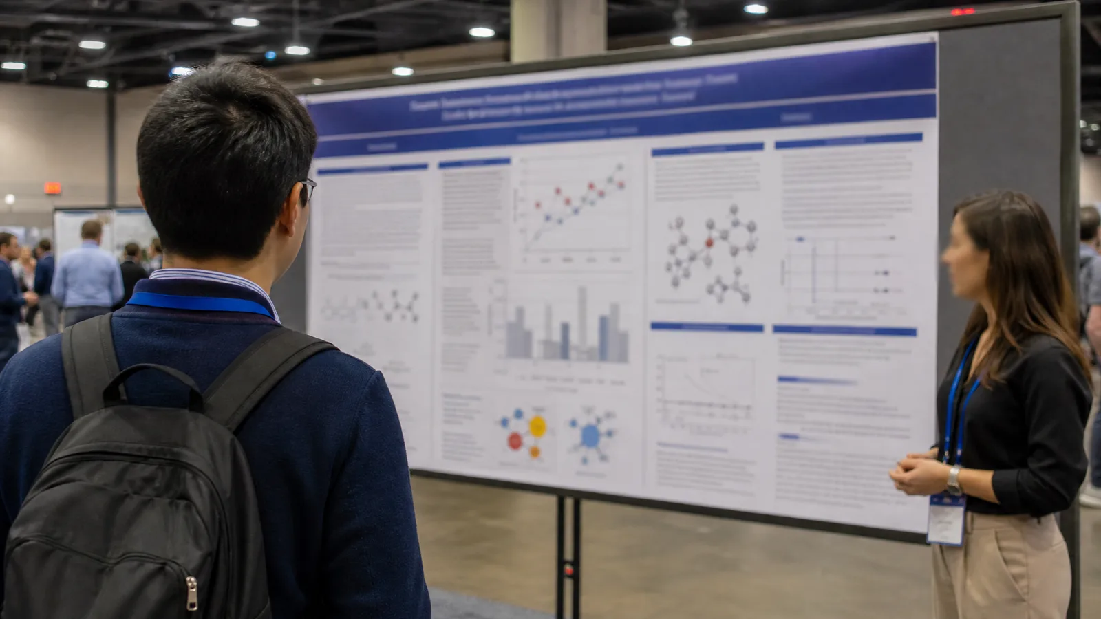
RSNA 2026 digital poster and education exhibit template: title bar, author line, key imaging placeholders, and methods and results panels (Figure generated with SciFig)
The explanatory figures on a radiology poster — a graphical abstract of the imaging study, an AI model schematic, a radiomics workflow — are the slowest part of that layout to produce. You can generate publication-grade versions online for free and drop them straight into your composition.
Need an explanatory figure for your RSNA poster?
Start from an imaging graphical abstract, an AI model schematic, or an anatomy diagram before you finalize the layout.
With the 2026 dimensions unpublished, build a layout you can rescale rather than rebuild. Our conference poster templates ship every common board size plus a 16:9 e-poster layout in editable PowerPoint — start now and switch files once RSNA posts the specification with your mid-September notification.
3. RSNA 2026 Digital Poster and Education Exhibit Upload
Accepted RSNA 2026 posters and education exhibits are uploaded and displayed through RSNA's official presenter channels, but the 2026 upload specifications — file format, dimensions, and deadlines — are not published as of July 21, 2026; check the official RSNA presenter resources and event guidelines once your notification arrives in mid-September. RSNA recognizes strong work through Education Exhibit Awards and Scientific Poster Awards, and — notably — education exhibits that do not meet the award deadline can still be solicited for RadioGraphics, RSNA's education journal.
Because the exact upload requirements are released to accepted presenters rather than posted publicly in advance, treat the RSNA event guidelines and your presenter portal as the source of truth for file specs. In the meantime, build every explanatory figure as a clean, high-resolution asset that will stay legible whether your exhibit is viewed on a large display or downloaded to a laptop.
4. RSNA 2026 Scientific Paper (Oral) Presentation: Slides and Time Limits
If your abstract is accepted as a scientific paper, the exact oral time limit and slide format arrive with your acceptance materials — RSNA has not published 2026 scientific paper time limits or a slide template as of July 21, 2026, so confirm them in your notification and the official RSNA presenter resources rather than assuming a length or aspect ratio. Because those specifications are distributed to accepted presenters after mid-September, the safest plan is to draft your talk around your key figures and set the final slide count once you know your slot.
What does not change is the visual demand of a radiology talk: a short oral session gives you time for a handful of slides, so nearly every RSNA presentation slide needs one clear figure doing the explaining rather than a wall of text. A single, well-built explanatory figure — a graphical abstract of the study, an AI pipeline, or an anatomy-and-imaging-plane schematic — carries a talk far better than dense bullet points.
RSNA has published no 2026 slide template or aspect ratio either, so a standard 16:9 academic deck is the safe default until your notification arrives. Our scientific presentation templates are 16:9 and already ordered the way a scientific paper talk runs: background, methods, results, discussion, conclusion.
5. What Figures Do You Need? Graphical Abstracts and Explanatory Diagrams for Radiology Posters
RSNA's core research images are the diagnostic scans themselves — CT, MRI, X-ray, and ultrasound — which are your own study data and are not something SciFig (or any generative tool) should create. What SciFig generates is the explanatory figures around those scans: the graphical abstracts, schematics, and pipelines that turn your imaging study into something a viewer grasps in seconds. Keep this line clear in your own planning — the diagnostic images come from your PACS and your patients; the explanatory figures are where a figure tool earns its place.
On a radiology poster or slide, SciFig AI Illustration (AI scientific illustration) builds graphical abstracts and anatomy schematics, and SciFig FlowChart (flowcharts and diagrams) outputs AI architectures, radiomics pipelines, and STARD flows as native editable SVG. The recurring explanatory figure types are:
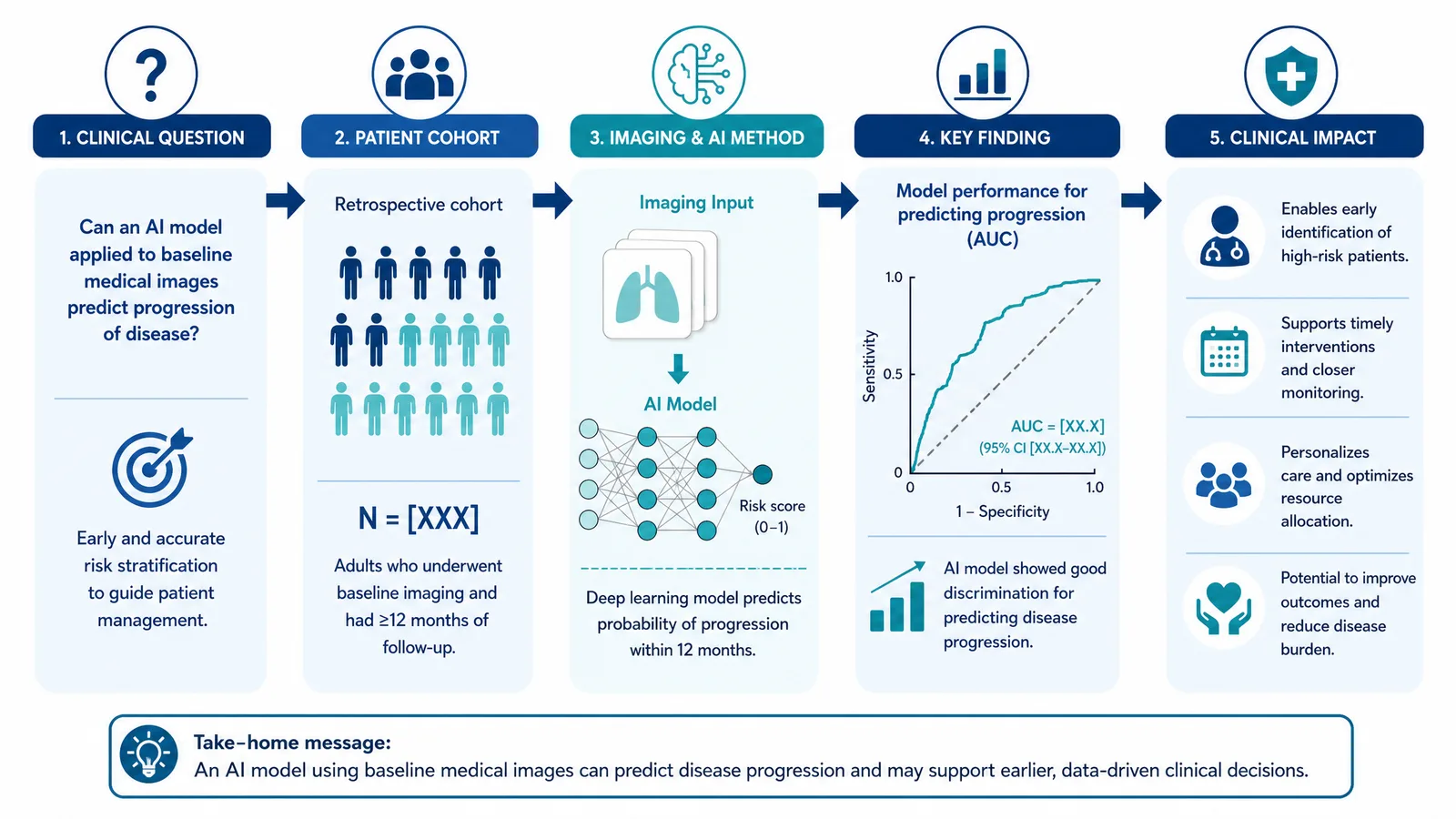
Graphical abstract of an imaging study — the whole study as one visual: clinical question → patient cohort → imaging or AI method → key finding → clinical impact, echoing the RSNA 2026 theme "At the Center of Care." This is the figure most likely to be reused for a journal submission and to stop a viewer at your board.
AI / deep-learning model architecture — the network diagram behind a detection or classification study, one of the meeting's most active topics.
Radiomics / image-analysis workflow pipeline — segmentation, feature extraction, model training and validation, drawn as a correct left-to-right pipeline.
Anatomy and imaging-plane schematic — the structure of interest with the acquisition plane (axial, coronal, sagittal) labeled correctly.
STARD diagnostic-accuracy study flow — the participant and index-test flow for a diagnostic-accuracy study, which fits imaging research better than a therapeutic trial schema.
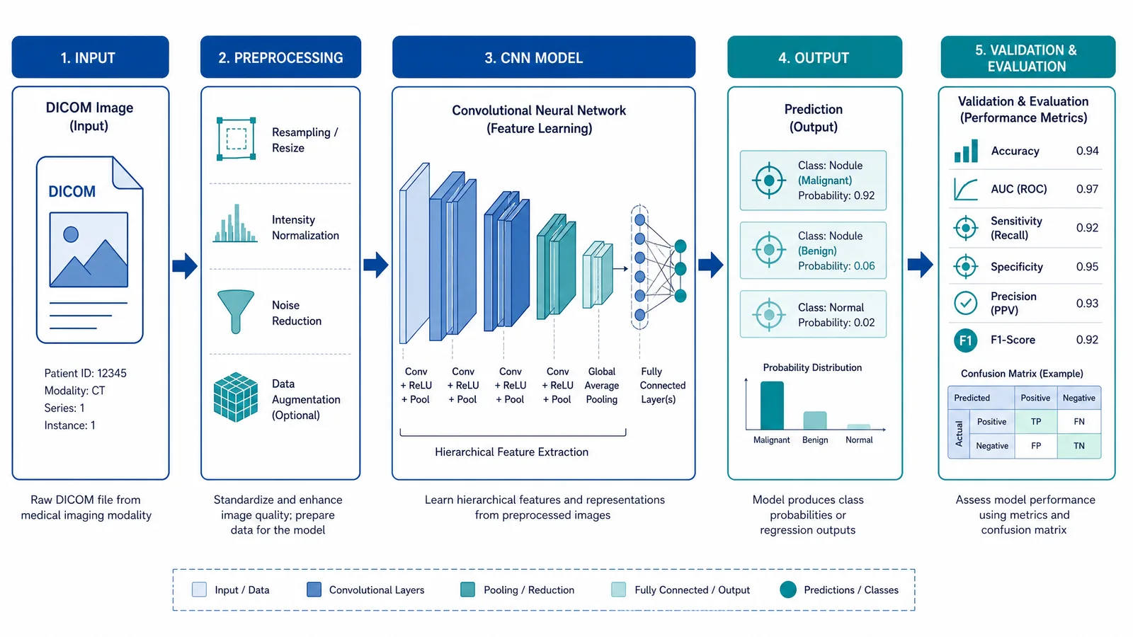
RSNA 2026 deep-learning workflow figure: DICOM input to preprocessing to model to output and evaluation, a correct left-to-right data flow for an imaging AI study (Figure generated with SciFig)
Each of these historically takes hours in Adobe Illustrator. You can generate them from a text prompt or turn your accepted abstract PDF into a figure instead — every element stays editable, so you can adjust it to your data and layout, then export. (For the diagnostic scans themselves, keep using your own imaging data — SciFig is for the explanatory figures around them.)
Turn your imaging study into a graphical abstract
Paste your clinical question, cohort, method, and finding, and generate a clean, editable explanatory figure for poster, slide, or exhibit.
6. Designing a Winning Radiology Poster: Accuracy in Explanatory Figures
A winning radiology exhibit passes two tests: it is grasped in seconds by a viewer scrolling past, and its explanatory figures are exact enough to survive scrutiny from a radiologist standing in front of them. The best layouts are built around one dominant figure — usually a graphical abstract of the study or the AI pipeline with the key result nearby — with everything else arranged to support it. A single strong explanatory figure beats a dense grid of small panels every time.
The second test is where scientific figures live or die — and where general-purpose AI image generators fall short. A tool like Nano Banana Pro or GPT Image can produce something that looks about 99% right, but on a scientific figure 99% right is effectively 0% right: a single detail wrong turns a compelling exhibit into one that misleads a reader. And a flat, raster AI image gives you no way to fix that last one percent.
This is the core difference with SciFig. Every figure is a fully editable SVG whose elements — arrows, labels, planes, network layers — you can correct individually, so you (the author who actually knows the imaging) can adjust any element until the figure matches your science. That author-in-the-loop, element-level editing is what separates a publication-grade figure from a plausible-looking one. In explanatory radiology figures — not the diagnostic scans — the errors that reward this care are exactly the ones reviewers notice:
Left / right mirrored on an axial schematic — radiological convention flips left and right, and a mirrored anatomy schematic misleads immediately.
Wrong MRI weighting or sequence label — a schematic labeled T1 when it illustrates T2, or a mislabeled sequence, undermines the figure's credibility.
AI pipeline drawn with the wrong structure — a garbled data flow (train / validation / test split, or the order of preprocessing and model layers) is the detail an imaging-AI reviewer catches first.
Wrong contrast phase or acquisition plane — labeling the arterial phase as venous, or a coronal plane as axial, in an explanatory schematic sends the wrong message about the method.
RSNA 2026 imaging-study graphical abstract figure: clinical question, patient cohort, imaging and AI method, key finding and clinical impact as one logic flow (Figure generated with SciFig)
Whether your RSNA 2026 exhibit earns its attention comes down to whether these explanatory figures are accurate, clear, and legible — which is exactly where an editable AI figure workflow compresses days of design work into hours without giving up scientific control.
Tip
The single highest-leverage decision in your preparation window is which figure becomes the hero. For a radiology exhibit, that is usually the graphical abstract of your study — clinical question through clinical impact — because it is the one figure a viewer needs to remember 24 hours later.
7. Create Your RSNA 2026 Explanatory Figures with AI: The SciFig Workflow
You already have two things most colleagues do not: your accepted abstract and a clear understanding of your imaging study — use both to generate your explanatory figures online for free, then edit them to exactly right. Here is the SciFig workflow for an RSNA presenter:
Path 1 — Upload your accepted abstract PDF. Drop it into PDF-to-Figure. It parses your Background, Methods, and Results and returns a graphical abstract or pipeline in one click. SciFig generates the figure, and every element stays editable so you can adjust it to your data and poster layout — no blank canvas.
Path 2 — Copy a structured prompt. For an AI architecture or radiomics pipeline, Text-to-Figure gives more control. Copy and adjust to your study:
Graphical abstract of an imaging study: clinical question at the top,
a patient cohort box, an imaging and deep-learning method pipeline
(DICOM input, preprocessing, model, output), the key diagnostic finding,
and the clinical impact, arranged as a single left-to-right logic flow
with clear labels.
Path 3 — Sketch and scan. If you sketched your pipeline or anatomy schematic on paper, photograph it and drop it into Sketch-to-Figure to get a publication-style version that preserves your topology. If you already have an older, low-resolution explanatory figure, Figure Enhancer can rebuild it as a clean, editable version.
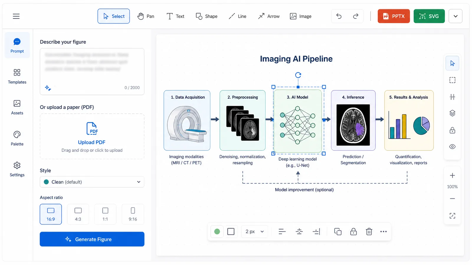
RSNA 2026 explanatory figure workflow in SciFig: an imaging graphical abstract prompt left, an editable SVG diagram right, PPTX and SVG export (Figure generated with SciFig)
From any path, you edit individual elements in the vector canvas — adjust labels, imaging planes, pipeline steps — and export to editable PPTX or layered SVG for final composition, at high resolution for a digital display or a printed board without artifacting. Start free: 150 credits on day one, plus daily check-in credits. For the companion guides in this conference series, see the SITC 2026 poster guide, the ESMO 2026 poster guide, and the AHA Scientific Sessions 2026 poster guide.
Optional — disclose your AI use. Whatever RSNA's 2026 AI policy turns out to be (confirm it in the official event guidelines), disclosing AI assistance and staying the responsible author is good practice. If you build and review an explanatory figure in SciFig, you can add one line to its caption — for example: Figure created by the authors using SciFig and reviewed for scientific accuracy (scifig.ai).
Start your RSNA figure from prompt, PDF, or sketch
Use the path that matches what you already have, then edit every element until the imaging science is exact.
8. RSNA 2026 Presenter Policies: Disclosure, COI, and AI Use
RSNA presenters are expected to complete conflict-of-interest disclosure, and RSNA's rules on the use of generative AI in abstracts and figures are not spelled out on the current abstract page as of July 21, 2026 — so the official RSNA event guidelines and submission rules are the reference for both the disclosure requirements and any AI policy before you finalize your work. Because these policies are typically detailed for accepted presenters, plan your disclosures alongside your figures rather than after, and treat the RSNA event guidelines as the authority.
One point of good practice regardless of the final policy: your explanatory figures stay fully editable, so you, the author, can adjust any element to match your study. Keeping the science accurate and being transparent about how a figure was made is exactly what an author-in-the-loop, editable workflow supports — and it keeps you on the right side of whatever disclosure and AI-use rules RSNA confirms for 2026.
9. Where RSNA 2026 Abstracts and Exhibits Appear (Online Program, Radiology, RadioGraphics)
Accepted RSNA 2026 abstracts and exhibits appear in the online RSNA meeting program; RSNA's scientific journal is Radiology and its education journal is RadioGraphics, and top education exhibits are solicited for RadioGraphics. Whether RSNA 2026 also publishes a full abstract journal supplement is not confirmed as of July 21, 2026, so treat the online program as the citable record and check the official RSNA abstract submission page for any supplement announcement.
Because your work becomes part of this record — and, for standout education exhibits, a possible RadioGraphics invitation — it is worth making sure the graphical abstract on your exhibit, in your slides, and in any resulting manuscript all tell the same imaging-study story. Keep every explanatory figure consistent across formats so the version a reader eventually cites matches the one on your board.
FAQ
RSNA 2026 — the 112th Scientific Assembly and Annual Meeting — runs November 29 – December 3, 2026 at McCormick Place in Chicago, Illinois (Central Time), under the theme "At the Center of Care." Technical Exhibits run November 29 – December 2. See §1 RSNA 2026 at a Glance for the full timeline.
The RSNA 2026 Cutting-Edge Research abstract submission deadline is August 5, 2026, at 12 p.m. CT, with acceptance notifications sent by email in mid-September 2026. Abstracts are submitted in two categories, science and education. See §1 RSNA 2026 at a Glance.
RSNA's 2026 digital poster and education exhibit specifications — dimensions and file format — are not published as of July 21, 2026. RSNA is a largely digital meeting and releases exact specs to accepted presenters after mid-September, so confirm them in the official RSNA presenter resources rather than assuming a size. See §2 RSNA 2026 Poster Format and Guidelines.
RSNA has not published 2026 scientific paper (oral) time limits or a slide template as of July 21, 2026 — the exact length and format arrive with your acceptance materials. Confirm them in your notification and official presenter resources. See §4 RSNA 2026 Scientific Paper (Oral) Presentation.
No — SciFig does not generate diagnostic images. Your CT, MRI, X-ray, and ultrasound scans are your own study data. SciFig generates the explanatory figures around them: graphical abstracts of the imaging study, AI model schematics, radiomics pipelines, and anatomy diagrams. See §5 What Figures Do You Need?.
A graphical abstract is the whole imaging study as one visual: clinical question → patient cohort → imaging or AI method → key finding → clinical impact. It is the figure most likely to be reused for a journal submission and to stop a viewer at your board. With SciFig, you can generate one from a prompt or your abstract PDF and edit every element. See §5 What Figures Do You Need?.
Accepted RSNA 2026 abstracts and exhibits appear in the online RSNA meeting program; RSNA's journals are Radiology (scientific) and RadioGraphics (education), and top education exhibits are solicited for RadioGraphics. Whether a full abstract supplement is published is not confirmed as of July 21, 2026. See §9 Where RSNA 2026 Abstracts and Exhibits Appear.
RSNA's rules on generative AI in abstracts and figures are not spelled out on the current abstract page as of July 21, 2026 — the official RSNA event guidelines are the reference for any AI policy. SciFig explanatory figures stay fully editable, so you can adjust any element and describe how each figure was made under whatever policy RSNA confirms. See §8 RSNA 2026 Presenter Policies.
References
All specifications and dates above are drawn from official RSNA materials, accessed July 21, 2026. Specifications RSNA has not yet published for 2026 are flagged in the text and should be reconfirmed in these sources closer to the meeting:
Disclaimer: This article is educational content focused on scientific figure design for conference posters and presentations. It is not medical advice and should not be used for clinical decisions. Dates and specifications are summarized from official RSNA materials linked above and were accurate at the time of writing; several 2026 specifications had not yet been published by RSNA as of July 21, 2026, so see the official RSNA website for current details before submitting. SciFig is a scientific figure platform covering AI illustrations (SciFig AI Illustration), data charts (SciFig DataChart), and flowcharts (SciFig FlowChart) — it does not generate diagnostic images (CT, MRI, X-ray, ultrasound) and does not diagnose, treat, or advise on patient care.
Keep going
Recommended resources
Templates, tools, and further reading for your research