SciFig

Free T-Cell Activation Diagram Generator

Arrange an antigen-presenting cell and a T cell with peptide–MHC/TCR recognition, B7/CD28 costimulation and a distinct cytokine signal.

Describe what you need in the box above.

Start with up to 150 free credits|No credit card required

Get up to 350 free credits on day one when you join through an invite.

3 figure engines · 500+ templatesEvery label editable · 60+ color schemesEditable PPTX · Layered SVG · 8K PNG / JPG

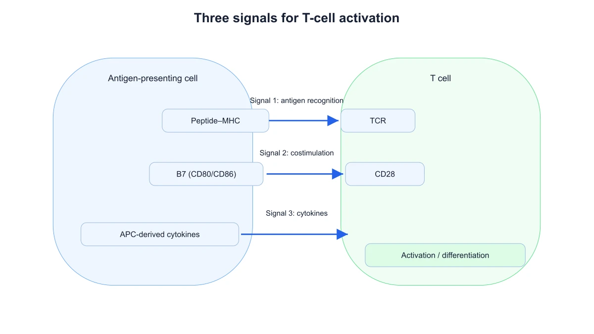
Three signals for T-cell activation

Choose a finite scientific preset, edit labels and export the result locally.

Diagram options
Scientific labels

Free T-Cell Activation Diagram Generator— templates & examples

Updated

T cell activation diagram tools

Free T-Cell Activation Diagram Generator

Arrange an antigen-presenting cell and a T cell with peptide–MHC/TCR recognition, B7/CD28 costimulation and a distinct cytokine signal.

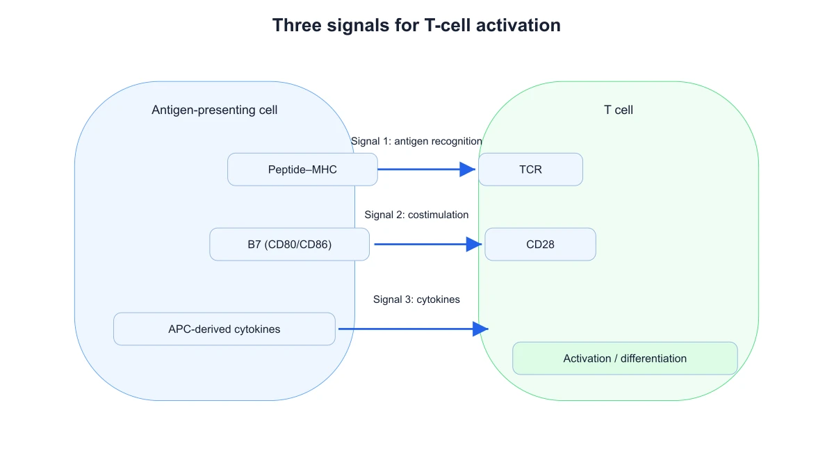
A T cell activation diagram shows antigen recognition, costimulation and cytokine signaling at the T-cell interface. Arrange an antigen-presenting cell and a T cell with peptide–MHC/TCR recognition, B7/CD28 costimulation and a distinct cytokine signal. The exported schematic retains the selected signaling labels.

APC and T-cell interface showing peptide–MHC/TCR recognition, B7/CD28 costimulation and a cytokine signal
  • Keep peptide–MHC on the APC
  • Locate B7 on the APC
  • Show a distinct cytokine signal
  • Edit cell-side labels

T Cell Activation Diagram Features

SciFig’s t cell activation diagram brings the inputs, diagram choices and exported result into one focused tool.

Show the antigen-recognition interface

Place peptide–MHC on the antigen-presenting cell opposite the T-cell receptor on the T cell. This paired layout gives the first signal a clear molecular and cellular location. Labels stay beside the corresponding side of the interface so the recognition partners are distinguishable.

  • Keep peptide–MHC on the APC
  • Keep TCR on the T cell
  • Identify the recognition signal
Show the antigen-recognition interface

Distinguish B7 and CD28 costimulation

Show B7 on the antigen-presenting cell paired with CD28 on the T cell. The costimulatory signal is separate from TCR recognition rather than another label for the same molecular contact. Use the signal control to focus a figure on recognition, costimulation or their relationship.

  • Locate B7 on the APC
  • Locate CD28 on the T cell
  • Separate costimulation from recognition
Distinguish B7 and CD28 costimulation

Add the cytokine signal

Use the cytokine signal to explain the third part of a three-signal activation model. Showing it independently lets the audience see that molecular recognition and costimulation are not the whole explanation. The diagram provides an organizing model without implying that all T-cell contexts have identical requirements.

  • Show a distinct cytokine signal
  • Toggle signal layers
  • Retain the three-signal distinction
Add the cytokine signal

Annotate the two cell sides

Use SciFig’s T cell activation diagram to change the cell and molecule labels to match your material, then export SVG or PNG. The figure retains the paired interface and visible signals. A compact layout is useful when the explanation needs molecular contacts in more detail than a general immune-response overview provides.

  • Edit cell-side labels
  • Label molecular partners
  • Export the activation interface
Annotate the two cell sides

How to Create the Figure

Use the t cell activation diagram controls below to prepare the figure from your selected mechanism and labels.

  1. Choose an activation-interface preset

    Start with the paired antigen-presenting-cell and T-cell interface. The preview locates peptide–MHC opposite TCR and places B7 opposite CD28. Choose an overview or focused example according to which contacts you want to introduce in your material.

  2. Choose costimulation and cytokine detail

    Use the costimulation and cytokine options to show the additional signal layers. Antigen recognition remains anchored to peptide–MHC and TCR. Add the visible layers that belong in your explanation while keeping the cell-side arrangement consistent.

  3. Edit the title and labels

    Enter a descriptive title and replace the node or structure labels with the terminology used in your document. The preset retains its arrangement while the visible names change. Keep molecular abbreviations consistent with the explanation beside the figure.

  4. Download the labeled figure

    Choose SVG for a scalable vector figure or PNG for a slide, handout or document. The exported diagram retains the selected view and labels. Keep the figure alongside the corresponding explanation so later wording changes can be reflected in another export.

T Cell Activation Diagram Uses

Use a labeled t cell activation diagram to connect the explanation in your material with a visible figure.

APC and T-cell interface showing peptide–MHC/TCR recognition, B7/CD28 costimulation and a cytokine signal

Introduce the three-signal model

Use the paired-cell figure to introduce antigen recognition, costimulation and cytokine signaling as distinct parts of an activation model. Add each layer to the explanation while keeping the same cell sides. Export the labeled figure for the accompanying lecture notes.

Explore this example
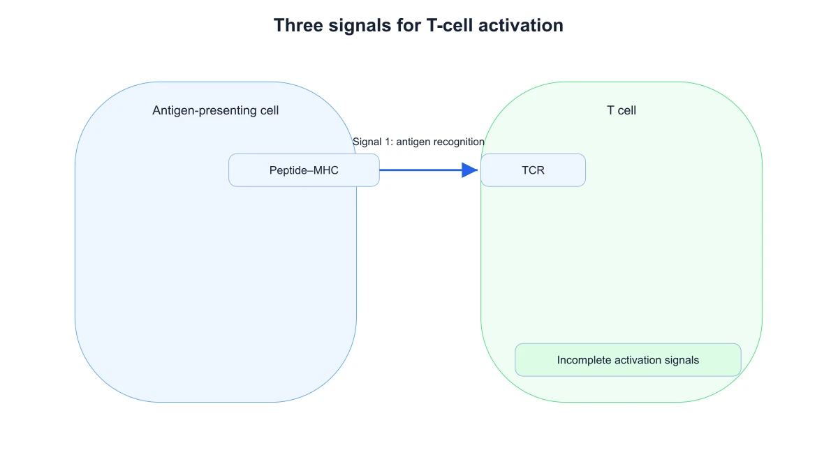
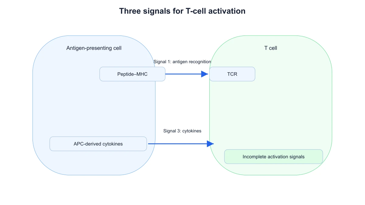
APC and T-cell interface showing antigen recognition alone and an incomplete-activation label

Explain peptide–MHC recognition

Focus on peptide–MHC opposite the TCR when the lesson introduces antigen recognition. Cell-side labels clarify which partner belongs on the antigen-presenting cell and which belongs on the T cell. The figure supplies a clear reference for the molecular names in the text.

Explore this example
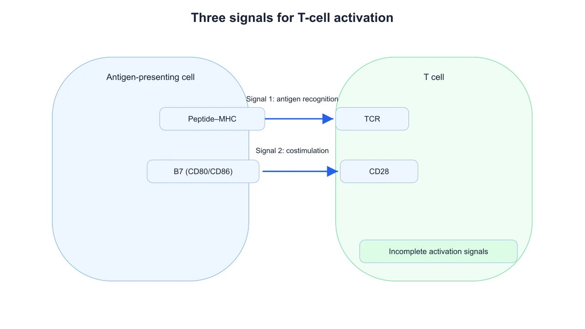
APC and T-cell interface showing antigen recognition and B7/CD28 costimulation without the cytokine signal

Discuss costimulation in a seminar

Show the B7/CD28 pair beside the recognition contact. The two interactions remain separate in the diagram, making it practical to discuss why costimulation is not simply another name for antigen binding. Keep the cell labels visible in the exported panel.

Explore this example
APC and T-cell interface showing antigen recognition and cytokine signaling without B7/CD28 costimulation

Connect cytokines with activation

Add the cytokine signal when moving from the cell-contact interface to the broader signaling environment. The separate signal helps organize that transition in a presentation. The schematic identifies roles and relationships without prescribing a clinical intervention or predicting an individual response.

Explore this example
Labeled APC and T-cell sides with all three activation-model signals displayed

Prepare a molecular-interface panel

Export SVG to place the T-cell interface beside a more detailed downstream signaling pathway. The interface panel identifies the cell-side partners, while the signaling panel explains subsequent molecular relationships. Keeping them distinct prevents a broad overview from becoming crowded with every pathway node.

Explore this example
APC and T-cell interface showing peptide–MHC/TCR recognition, B7/CD28 costimulation and a cytokine signal

Extend an immune-response overview

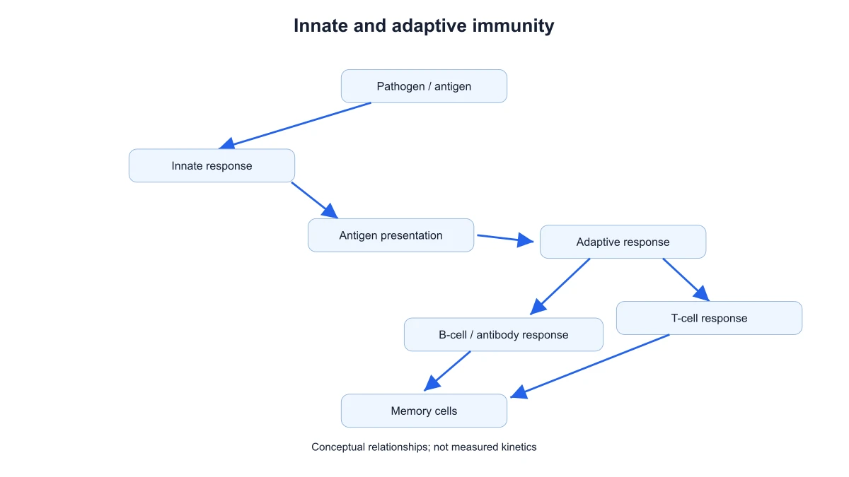
Use the dedicated activation figure after a general innate-and-adaptive overview. The larger overview establishes context; this panel explains peptide–MHC/TCR recognition and costimulation at the contact between cells. A consistent set of labels helps the audience move between the two levels.

Explore this example
How Researchers Use SciFig

My seminar figure needed the receptor partners on the correct sides of the cell contact. The T cell activation diagram placed peptide–MHC opposite TCR and B7 opposite CD28. I edited the labels and exported SVG, which gave the audience a reference for recognition and costimulation before the downstream signaling discussion.

Laura M.

Cellular immunology doctoral researcher, University of Lausanne

Free tools

A tool for every scientific figure

120 focused generators for the scientific figure you need — each opens in the right workspace with a matching example.

Illustrations, structures and concept diagrams

60 tools
Chemistry Structure Generator example figureChemistry Structure GeneratorGraphical Abstract Maker example figureGraphical Abstract MakerFree Body Diagram Generator example figureFree Body Diagram GeneratorCircuit Diagram Maker example figureCircuit Diagram MakerAI Scientific Image Generator example figureAI Scientific Image GeneratorEditable 96-Well Plate example figureEditable 96-Well PlateLewis Dot Structure Generator example figureLewis Dot Structure GeneratorDNA Structure Diagram Generator example figureDNA Structure Diagram GeneratorWiring Diagram Maker example figureWiring Diagram MakerP&ID Diagram Generator example figureP&ID Diagram GeneratorScientific Poster Generator example figureScientific Poster GeneratorCladogram Maker example figureCladogram MakerFood Web Maker example figureFood Web MakerMedical Illustration Generator example figureMedical Illustration GeneratorPCR Diagram Generator example figurePCR Diagram GeneratorPPT Text to Visual Converter example figurePPT Text to Visual ConverterMitosis Diagram Generator example figureMitosis Diagram GeneratorAI Infographic Generator example figureAI Infographic GeneratorAnatomical Drawing Generator example figureAnatomical Drawing GeneratorAnimal and Plant Cell Comparison example figureAnimal and Plant Cell ComparisonAnimal Cell Diagram Generator example figureAnimal Cell Diagram GeneratorAntibody Structure Diagram Generator example figureAntibody Structure Diagram GeneratorApoptosis Diagram Generator example figureApoptosis Diagram GeneratorBacteria Diagram Generator example figureBacteria Diagram GeneratorBlueprint Maker example figureBlueprint MakerBohr Model Maker example figureBohr Model MakerCalvin Cycle Diagram Generator example figureCalvin Cycle Diagram GeneratorCarbon Cycle Diagram Generator example figureCarbon Cycle Diagram GeneratorCell Cycle Diagram Generator example figureCell Cycle Diagram GeneratorCellular Respiration Diagram Generator example figureCellular Respiration Diagram GeneratorComplement System Diagram Generator example figureComplement System Diagram GeneratorCRISPR-Cas9 Diagram Generator example figureCRISPR-Cas9 Diagram GeneratorDNA Replication Diagram Generator example figureDNA Replication Diagram GeneratorElectron Configuration Diagram Generator example figureElectron Configuration Diagram GeneratorElectron Transport Chain Diagram Generator example figureElectron Transport Chain Diagram GeneratorGene Regulation Diagram Generator example figureGene Regulation Diagram GeneratorGlycolysis Diagram Generator example figureGlycolysis Diagram GeneratorImage to Editable SVG Converter example figureImage to Editable SVG ConverterImmune Response Diagram Generator example figureImmune Response Diagram GeneratorKrebs Cycle Diagram Generator example figureKrebs Cycle Diagram GeneratorLab Diagram Maker example figureLab Diagram MakerMeiosis Diagram Generator example figureMeiosis Diagram GeneratorMolecular Orbital Diagram Generator example figureMolecular Orbital Diagram GeneratorNitrogen Cycle Diagram Generator example figureNitrogen Cycle Diagram GeneratorPedigree Chart Maker example figurePedigree Chart MakerPhospholipid Diagram Generator example figurePhospholipid Diagram GeneratorPhosphorus Cycle Diagram Generator example figurePhosphorus Cycle Diagram GeneratorPhotosynthesis Diagram Generator example figurePhotosynthesis Diagram GeneratorPhylogenetic Tree Maker example figurePhylogenetic Tree MakerPhysics Vector Diagram Maker example figurePhysics Vector Diagram MakerPlant Cell Diagram Generator example figurePlant Cell Diagram GeneratorProtein Synthesis Diagram Generator example figureProtein Synthesis Diagram GeneratorPunnett Square Maker example figurePunnett Square MakerRay Diagram Maker example figureRay Diagram MakerScientific Color Palette Generator example figureScientific Color Palette GeneratorSignaling Pathway Diagram Generator example figureSignaling Pathway Diagram GeneratorSingle Line Diagram Generator example figureSingle Line Diagram GeneratorT-Cell Activation Diagram Generator example figureT-Cell Activation Diagram GeneratorWater Cycle Diagram Generator example figureWater Cycle Diagram GeneratorWestern Blot Diagram Generator example figureWestern Blot Diagram Generator
T Cell Activation Diagram Questions

1.

2.

3.

4.

5.

6.

Call to action background

T cell activation diagram

Create a t cell activation diagram

Create a t cell activation diagram

Free to start · No credit card required · Built for researchers