
Pseudomonas Aeruginosa
A labeled, publication-ready figure of the Pseudomonas aeruginosa cell — its gram-negative envelope, flagellum, pili, and key virulence factors.
Updated

Start with up to 150 free credits|No credit card required
Get up to 350 free credits on day one when you join through an invite.
What is Pseudomonas Aeruginosa?
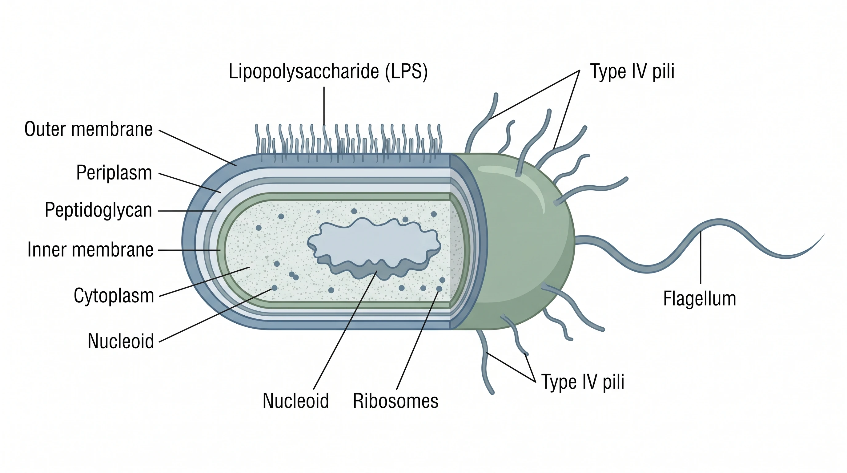
Pseudomonas aeruginosa is a rod-shaped, gram-negative bacterium known for its single polar flagellum, type IV pili, and many virulence factors. A labeled structure diagram shows its outer membrane and lipopolysaccharide, periplasm, cytoplasm, flagellum, pili, and secreted toxins, helping explain its motility, biofilms, and antibiotic resistance. With SciFig you describe the cell in plain language and generate a clean, labeled Pseudomonas aeruginosa figure for your paper, poster, or slides.
Why one labeled reference cell is worth drawing properly
- Intrinsic resistance cannot be explained without the envelope. A reviewer reading an MIC result expects the permeability and efflux story to be drawn, not asserted.
- One canonical cell keeps every panel in a paper consistent — same pole, same flagellum, same colors for inner and outer membrane across figures.
- It is where clinical and molecular readers meet: the same drawing carries the ICU infection sites and the drug targets.
- Onboarding: a new lab member needs to understand why OprD loss matters before running a carbapenem assay, and a labeled figure does that faster than a paper.
- Grant reviewers judge whether a proposed target is reachable at all — a periplasmic target and a secreted target are very different bets, and the figure makes the difference obvious.
- Figures drawn from memory routinely add peritrichous flagella or a thick peptidoglycan layer; starting from a reviewed template removes that whole class of error.
Key components to label
- Cell body and single polar flagellum — swimming motility and chemotaxis
- Type IV pili — twitching motility, surface adhesion, and natural competence
- Outer membrane with lipopolysaccharide — lipid A, core oligosaccharide, and O-antigen
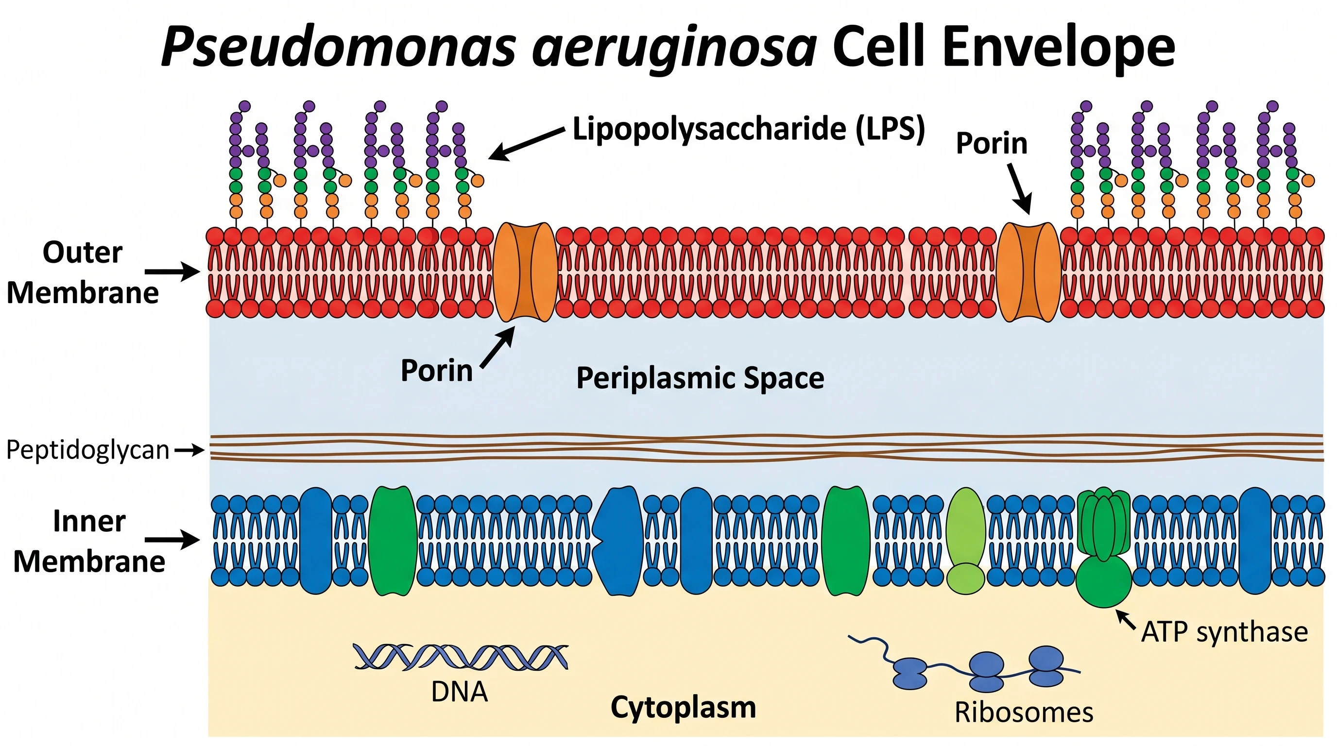
- Porins and efflux — OprF and OprD channels, MexAB-OprM and related RND pumps
- Periplasm and thin peptidoglycan layer, with inducible AmpC β-lactamase
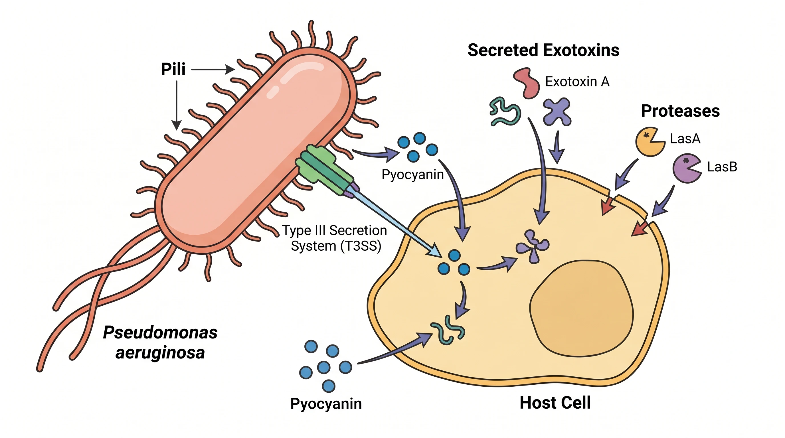
- Secretion systems — T3SS injectisome delivering ExoU/ExoS/ExoT/ExoY, and T2SS products elastase LasB, exotoxin A, and phospholipase C
- Diffusible products and their regulators — pyocyanin, pyoverdine, pyochelin, and the las/rhl/pqs quorum-sensing circuits; add the Psl/Pel/alginate matrix when the biofilm state is shown
Where researchers use this figure
- Graphical abstracts for antimicrobial-resistance and antibiotic-discovery papers
- Reviews of biofilm biology and chronic cystic-fibrosis airway infection
- Grant background figures locating a proposed drug target in the envelope or the secretome
- Clinical microbiology and infectious-disease teaching on non-fermenting gram-negative rods
- Infection-control training covering ventilator-associated pneumonia, burn wounds, keratitis, and catheter-related infection
- Thesis introductions and lab SOPs that need one canonical labeled cell everyone works from
Everything you need in a publication-ready P. aeruginosa cell figure
Get the morphology and surface appendages right
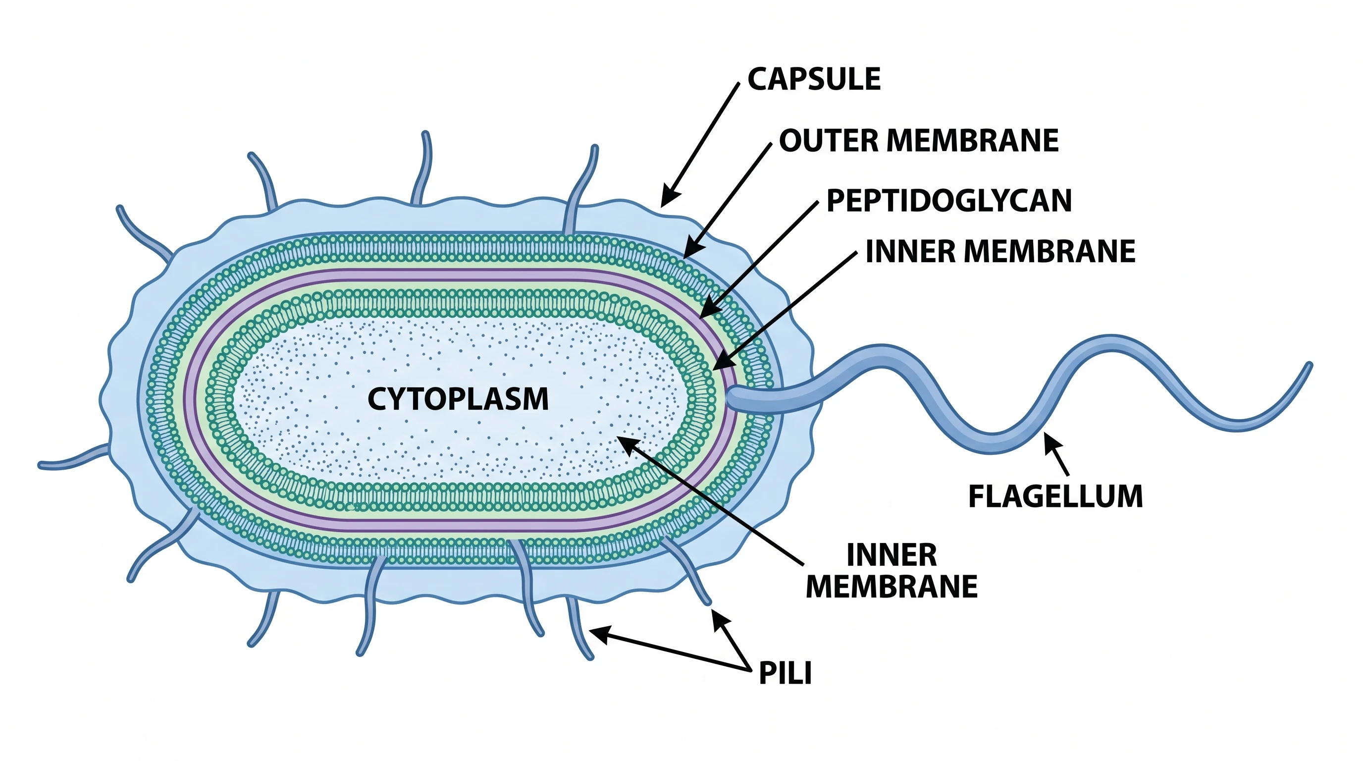
A rod roughly 0.5–0.8 µm wide and 1.5–3 µm long, with a single polar flagellum for swimming and chemotaxis and type IV pili at the same pole for twitching motility, surface attachment, and DNA uptake. Two appendages, one pole, different jobs — drawings that scatter peritrichous flagella around the cell are describing a different organism. A scale bar makes the panel usable in a methods figure.
Label the gram-negative envelope layer by layer
Inner membrane, thin peptidoglycan in the periplasm, outer membrane with lipopolysaccharide facing out — lipid A, core, and O-antigen. The porins are where the pharmacology lives: OprD admits carbapenems and resistance follows its loss, while low outer-membrane permeability plus MexAB-OprM efflux explains why this envelope is intrinsically hard for antibiotics to cross. Inducible AmpC β-lactamase sits in the same periplasmic space and belongs on the panel.
Group virulence factors by how they leave the cell
The type III secretion system injects ExoU, ExoS, ExoT, and ExoY straight into host cells on contact; the type II system releases elastase LasB, exotoxin A, and phospholipase C into the medium; pyocyanin and the siderophores pyoverdine and pyochelin diffuse freely. Grouping by secretion route rather than by alphabet gives the panel a logic, and leaves an obvious place for the las, rhl, and pqs quorum-sensing circuits that switch them on.
Show the biofilm as a matrix, not a blob
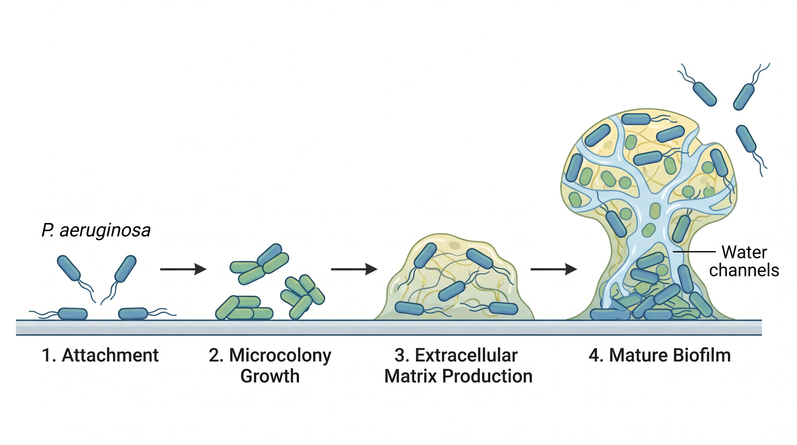
Attachment, microcolony, mature biofilm, dispersal — four stages, each with its own labels. The matrix is Psl, Pel, and alginate, held together under c-di-GMP control; mucoid conversion after mucA mutation is what marks chronic cystic-fibrosis airway isolates. Tolerance in the biofilm state comes from slow growth and limited matrix penetration, not from a new resistance gene, and the figure should not blur the two.
Pseudomonas Aeruginosa— templates & examples
How to make Pseudomonas Aeruginosa
Describe your figure
Tell SciFig what to draw in plain language — no design tools required.
Generate with SciFig
Get a clean, publication-ready figure that matches your description in seconds.
Edit & export
Vectorize it into editable SVG, relabel everything, and export for your paper, poster, or slides.
Related searches
- pseudomonas aeruginosa diagram
- pseudomonas aeruginosa structure
- pseudomonas cell diagram
- pseudomonas virulence factors
- gram negative bacteria structure
- pseudomonas aeruginosa labeled
Make figures for
Related pages
Microbiology Templates
Browse more Microbiology templates in SciFig's library.
All Templates
Browse every template page in SciFig's library.
Zoonotic Transmission of Salmonella
A publication-ready figure mapping how Salmonella moves from animal reservoirs through food, water, and the environment to infect humans.
Conjugation Transformation Transduction
One clear figure comparing the three mechanisms of bacterial horizontal gene transfer: conjugation, transformation, and transduction.
Graphical Abstract Maker
Summarize your study in one panel
Scientific Poster Generator
Design a conference research poster
AI Scientific Image Generator
Generate research figures from text
Conceptual Framework Generator
Map variables and their relationships
Frequently Asked Questions
Common questions about Pseudomonas Aeruginosa.

Ready to publish?
Make your own Pseudomonas Aeruginosa in minutes.
Start for freeFree to start · No credit card required · Built for researchers