
Conjugation Transformation Transduction
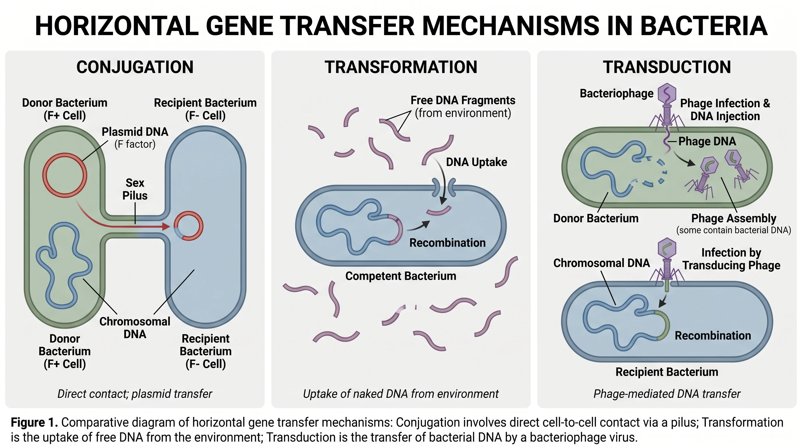
One clear figure comparing the three mechanisms of bacterial horizontal gene transfer: conjugation, transformation, and transduction.
Updated

Start with up to 150 free credits|No credit card required
Get up to 350 free credits on day one when you join through an invite.
What is Conjugation Transformation Transduction?
Conjugation, transformation, and transduction are the three mechanisms of horizontal gene transfer in bacteria. Conjugation moves DNA directly between cells through a pilus; transformation is the uptake of free DNA from the environment; and transduction transfers DNA between bacteria using a bacteriophage. A side-by-side diagram makes the differences easy to compare. With SciFig you describe the three mechanisms in plain language and generate a labeled figure for lectures or papers.
Why one comparative figure beats three separate ones
- Students and reviewers alike collapse transformation and transduction; a single aligned figure is the cheapest correction available.
- Resistance papers have to state which mechanism their data support — a mating assay, a transformation frequency, and a phage-mediated transfer experiment are not interchangeable evidence.
- The mechanism determines the barrier. Restriction–modification, CRISPR-Cas, and surface exclusion act at different points, and the figure is where you place them.
- Methods sections lean on it to justify controls: a DNase-treated arm rules out uptake of free DNA; a filter-mating arm rules out contact-independent transfer.
- Reuse across a thesis or lecture series: one consistent set of icons saves the reader from re-learning the notation in every chapter.
- Biosafety and engineered-strain reviews ask explicitly which transfer routes are open, and expect a drawn answer.
Key components to label
- Donor cell carrying a conjugative plasmid — tra operon, oriT, and the mating-pair-forming type IV secretion system
- Conjugative pilus and the transferred single strand, with rolling-circle replication maintaining the donor copy
- Extracellular DNA released by lysed cells — the substrate for transformation
- Competence machinery in the recipient — pseudopilus, ComEA DNA receptor, ComEC translocase, and degradation of the non-transported strand
- Bacteriophage life cycle — adsorption to a surface receptor, injection, and the branch between lytic replication and lysogenic integration
- Packaging of host DNA — a random fragment in generalised transduction, prophage-flanking genes in specialised transduction
- The shared endpoint — RecA-mediated homologous recombination into the chromosome, or autonomous replication if the element is a plasmid or ICE, plus the defense systems that block both
Where researchers use this figure
- Microbiology and genetics lecture slides, where the three mechanisms are always taught together
- Review articles and thesis introductions on the spread of antimicrobial-resistance genes
- Grant background sections arguing that plasmid-mediated resistance drives a clinical problem
- Method schematics for mating assays, competence protocols, and phage-transfer experiments
- Conference posters and graphical abstracts that must be recognisable from two meters away
- Biosafety and engineered-organism risk documents that have to state which transfer routes are plausible
Everything you need in a publication-ready horizontal gene transfer figure
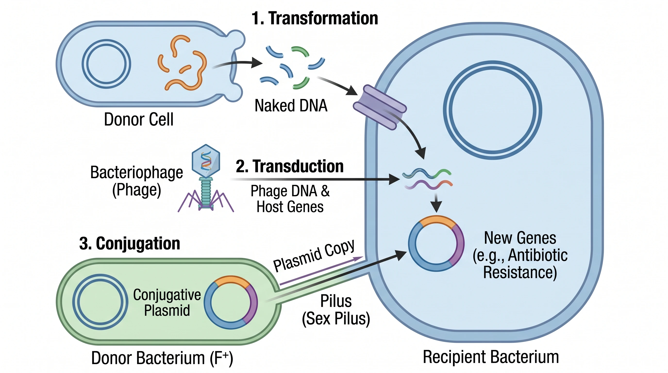
Put the three mechanisms on one comparable grid
Side by side, the distinction is easy to hold: conjugation needs cell-to-cell contact, transformation needs no donor cell at all, and transduction needs a phage. Aligning the panels so the donor, the DNA, the vehicle, and the recipient sit in the same position in each row is what makes the comparison readable. Each row then ends in the same place — recombination or plasmid maintenance.
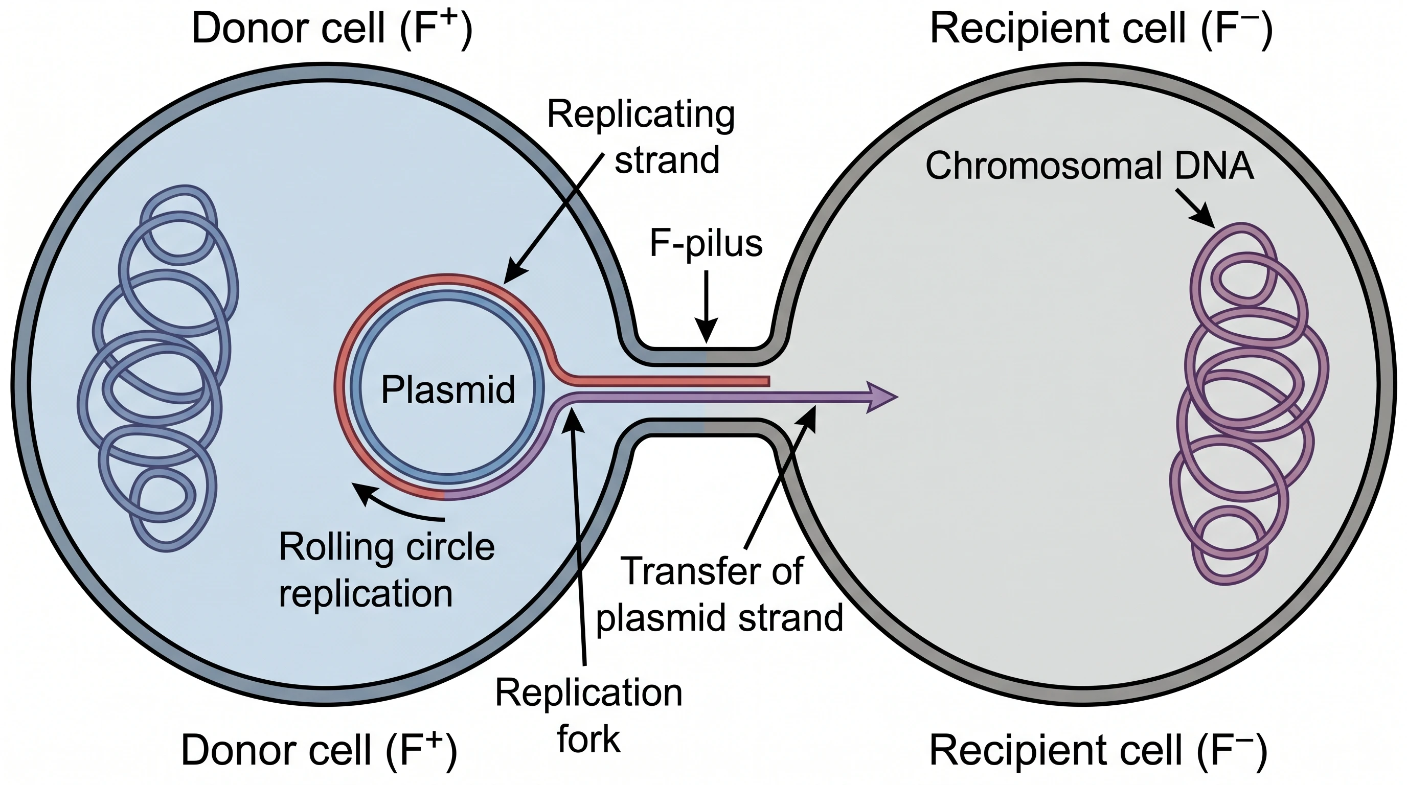
Draw conjugation as a single-strand transfer event
Most textbook drawings show a whole plasmid sliding down a pilus, which is wrong. The relaxase nicks the plasmid at oriT, one strand is transferred through a type IV secretion system while rolling-circle replication regenerates the donor copy, and the recipient synthesises the complementary strand. Labeling the tra genes, oriT, and the mating-pair contact keeps the panel accurate for a resistance-plasmid audience.
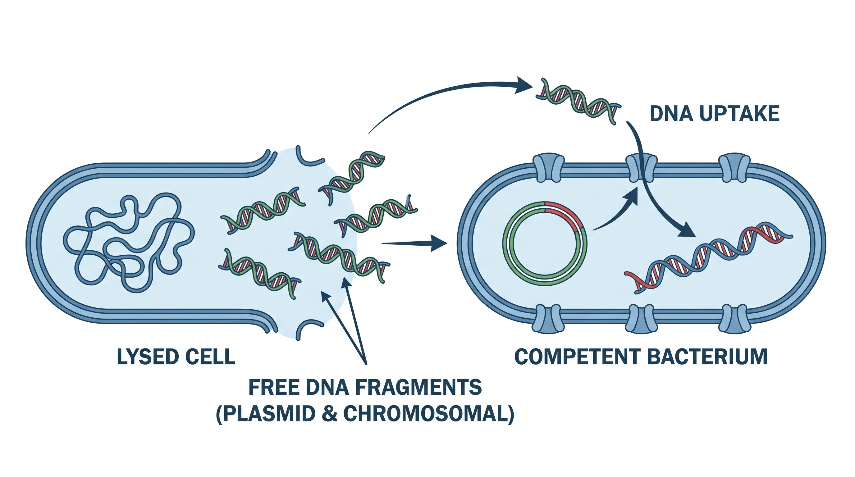
Show competence, not just floating DNA
Uptake of free DNA is an active, regulated process: competence genes switch on, a pseudopilus and the ComEA receptor bind extracellular DNA, ComEC threads a single strand across the membrane while the other strand is degraded, and RecA drives homologous recombination into the chromosome. Species with uptake-sequence bias — Neisseria DUS, Haemophilus USS — deserve a note, because they discriminate against foreign DNA.
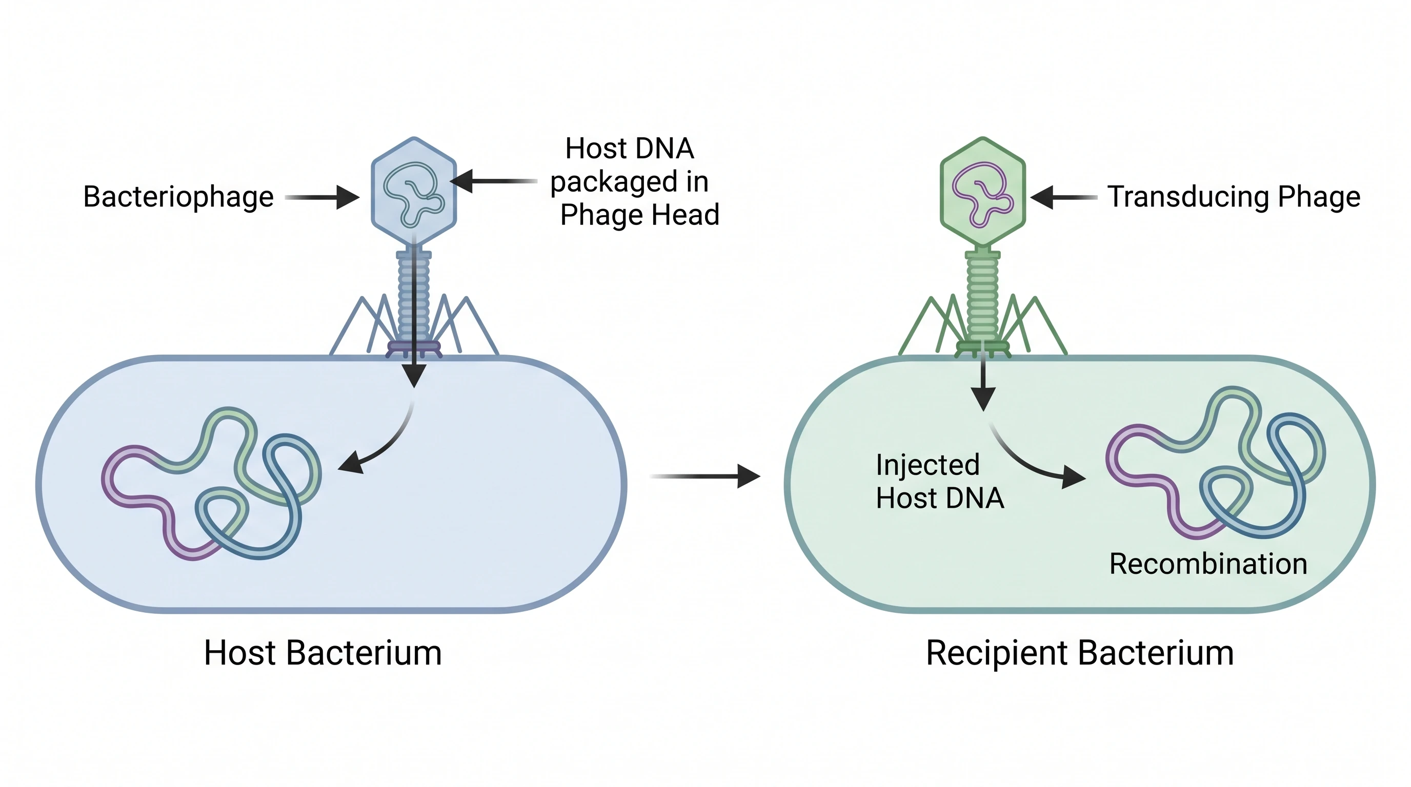
Split transduction into generalised and specialised
These are two different figures. Generalised transduction is a packaging error — headful packaging in P1 or P22 captures a random fragment of host chromosome instead of phage DNA. Specialised transduction follows imprecise excision of an integrated prophage such as lambda, so only genes flanking the integration site travel, classically gal and bio. Drawing both, with the lytic and lysogenic cycles labeled, prevents the most common error.
Conjugation Transformation Transduction— templates & examples
How to make Conjugation Transformation Transduction
Describe your figure
Tell SciFig what to draw in plain language — no design tools required.
Generate with SciFig
Get a clean, publication-ready figure that matches your description in seconds.
Edit & export
Vectorize it into editable SVG, relabel everything, and export for your paper, poster, or slides.
Related searches
- bacterial conjugation transformation transduction
- transformation vs transduction
- horizontal gene transfer diagram
- bacterial gene transfer mechanisms
- what is bacterial conjugation
- bacteriophage transduction
Make figures for
Related pages
Microbiology Templates
Browse more Microbiology templates in SciFig's library.
All Templates
Browse every template page in SciFig's library.
Zoonotic Transmission of Salmonella
A publication-ready figure mapping how Salmonella moves from animal reservoirs through food, water, and the environment to infect humans.
Pseudomonas Aeruginosa
A labeled, publication-ready figure of the Pseudomonas aeruginosa cell — its gram-negative envelope, flagellum, pili, and key virulence factors.
Graphical Abstract Maker
Summarize your study in one panel
Scientific Poster Generator
Design a conference research poster
AI Scientific Image Generator
Generate research figures from text
Conceptual Framework Generator
Map variables and their relationships
Frequently Asked Questions
Common questions about Conjugation Transformation Transduction.

Ready to publish?
Make your own Conjugation Transformation Transduction in minutes.
Start for freeFree to start · No credit card required · Built for researchers