
Drugs Acting on Various Pathophysiological Pathways in Ulcerative Colitis
A publication-ready figure showing where each class of ulcerative colitis drug acts on the inflamed colon's pathophysiological pathways.
Updated

Start with up to 150 free credits|No credit card required
Get up to 350 free credits on day one when you join through an invite.
What is Drugs Acting on Various Pathophysiological Pathways in Ulcerative Colitis?
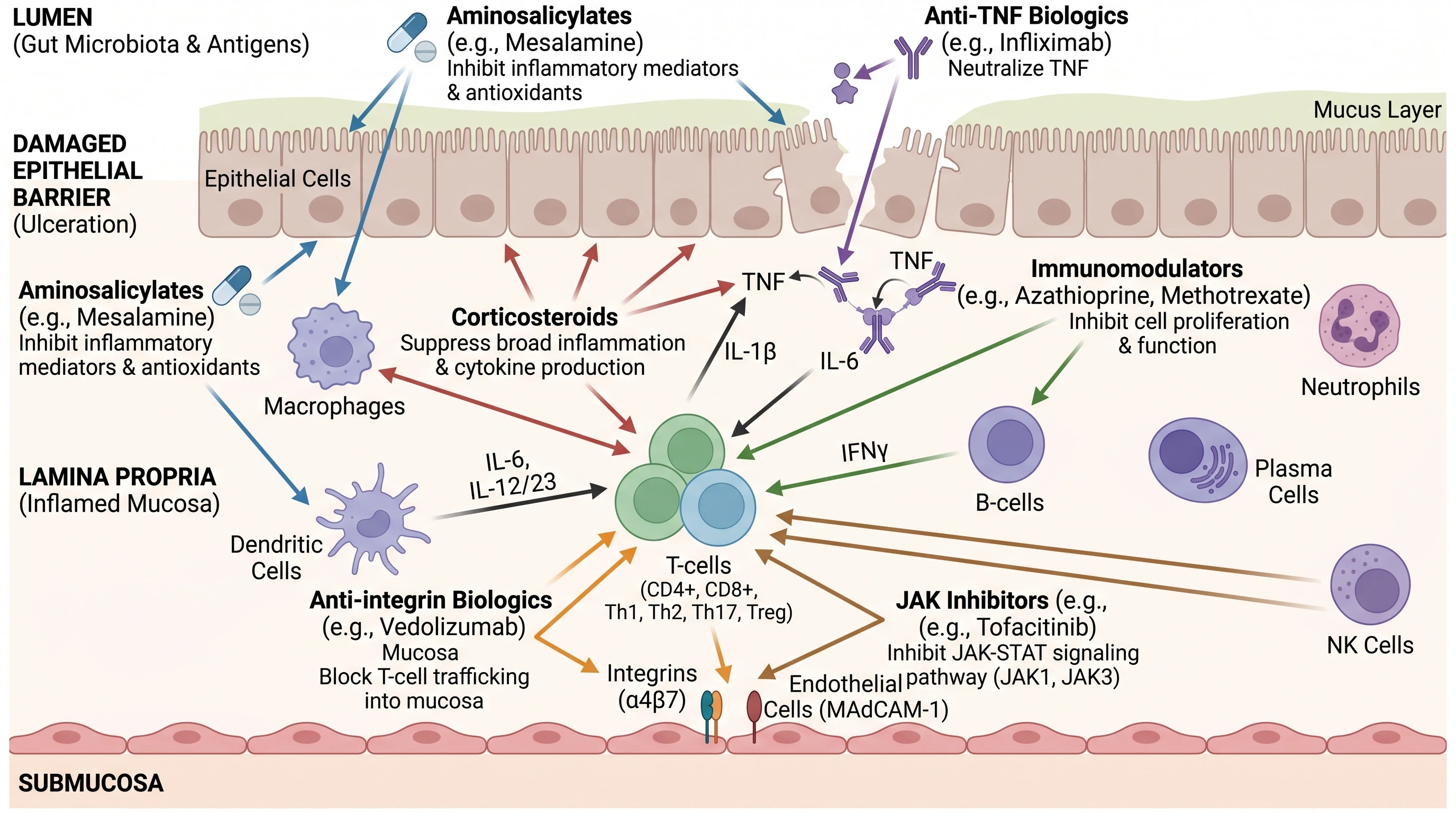
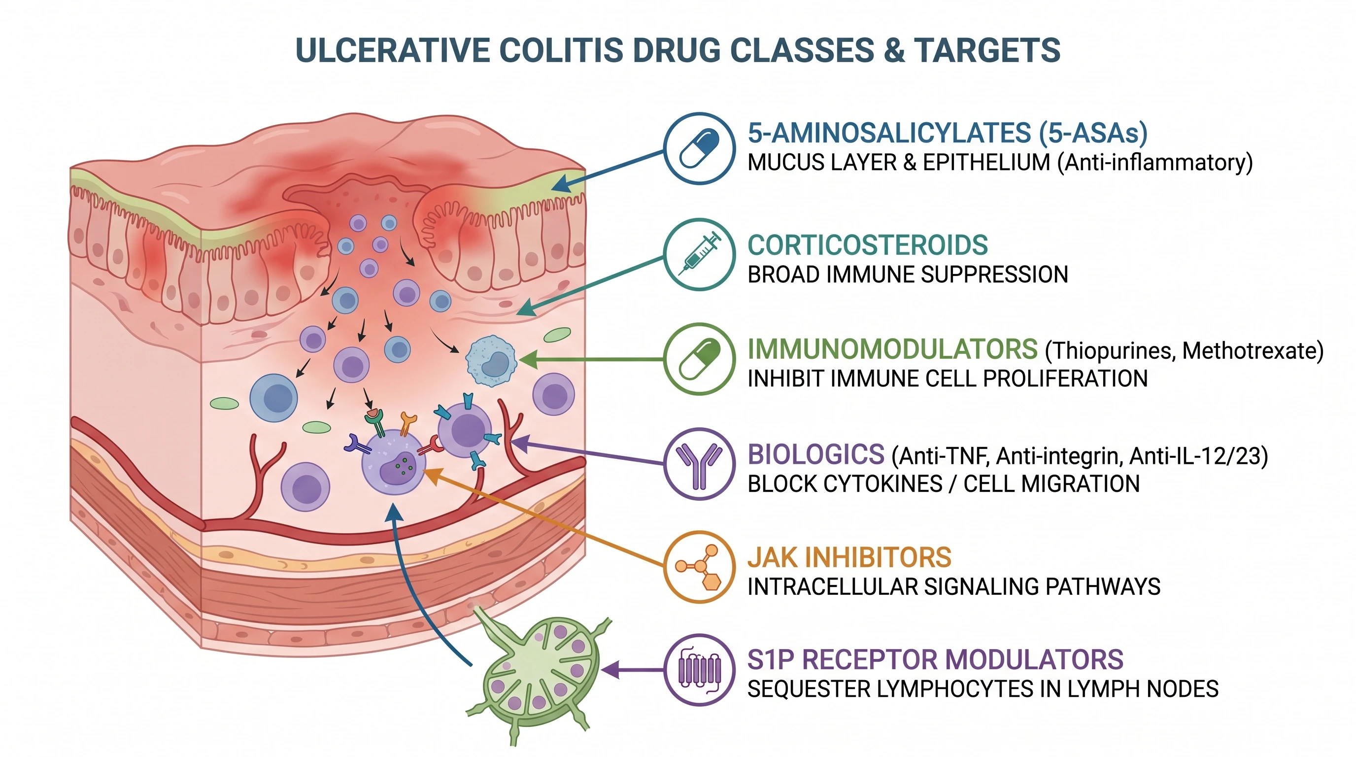
Drugs for ulcerative colitis act on several pathophysiological pathways that drive inflammation in the colon. Aminosalicylates work on the mucosa, corticosteroids broadly suppress inflammation, and immunomodulators and biologics target molecules such as TNF, integrins, and JAK signaling. A labeled pathway figure maps each drug class to its target on the colon mucosa. With SciFig you describe the targets and generate a clean, editable diagram for your paper or slides.
Why a mechanism map earns its place in the paper
- A treatment algorithm without a mechanism map is just a list. Positioning a new agent requires showing which targets are already occupied and at what level they act.
- Reviewers expect the mechanism and the level of action to agree — a JAK inhibitor drawn outside the cell is an immediate red flag.
- Trainees learn class distinctions faster from compartments than from tables: luminal and topical, broadly systemic, trafficking, intracellular.
- Trial protocols and grant backgrounds must state where an investigational drug sits relative to approved targets, and a figure states it in one glance.
- Safety follows mechanism. Gut-selective integrin blockade, broad JAK inhibition, and lymphocyte sequestration carry different risks, and the figure is where that argument starts.
- Ulcerative colitis and Crohn's disease share many agents but not all; a drawn colon keeps the discussion honest about which segment the evidence actually covers.
Key components to label
- Mucosal compartments — lumen and microbiota, mucus layer and goblet cells, epithelium, lamina propria, submucosal venule
- Aminosalicylates at the epithelial surface — mesalamine, sulfasalazine, balsalazide — with the delivery system named alongside the drug
- Corticosteroids — glucocorticoid receptor transrepression of NF-κB and AP-1; budesonide MMX flagged as high first-pass metabolism and induction only
- Thiopurines — azathioprine and 6-mercaptopurine converted to 6-thioguanine nucleotides, with TPMT and NUDT15 status noted
- Cytokine-neutralising biologics — anti-TNF (infliximab, adalimumab, golimumab) and the IL-12/23 p40 and IL-23 p19 antibodies (ustekinumab, mirikizumab)
- Leukocyte-trafficking agents at the vessel wall — vedolizumab against the α4β7–MAdCAM-1 interaction, and S1P modulators sequestering lymphocytes in lymph nodes
- Intracellular small molecules — JAK inhibitors acting on the JAK–STAT axis inside the target cell
Where researchers use this figure
- Review articles and graphical abstracts on IBD therapeutics
- Grant and trial-protocol background sections positioning an investigational agent against existing targets
- Gastroenterology fellowship and clinical pharmacology teaching slides
- Guideline and treatment-algorithm figures that pair positioning advice with mechanism
- Thesis introductions and journal clubs comparing biologics with oral small molecules
- Conference posters where the mechanism has to be legible without reading the abstract
Everything you need in a publication-ready UC drug-target figure
Anchor every drug class to a mucosal compartment
The figure only works if the anatomy is drawn first: lumen and microbiota, the epithelial barrier with its depleted mucus layer, the lamina propria packed with macrophages, dendritic cells, and T cells, and the submucosal venule where circulating lymphocytes are recruited. Each drug class then has one honest home. A reader should be able to point at a compartment and name the agents acting there.
Show 5-ASA acting topically, not systemically
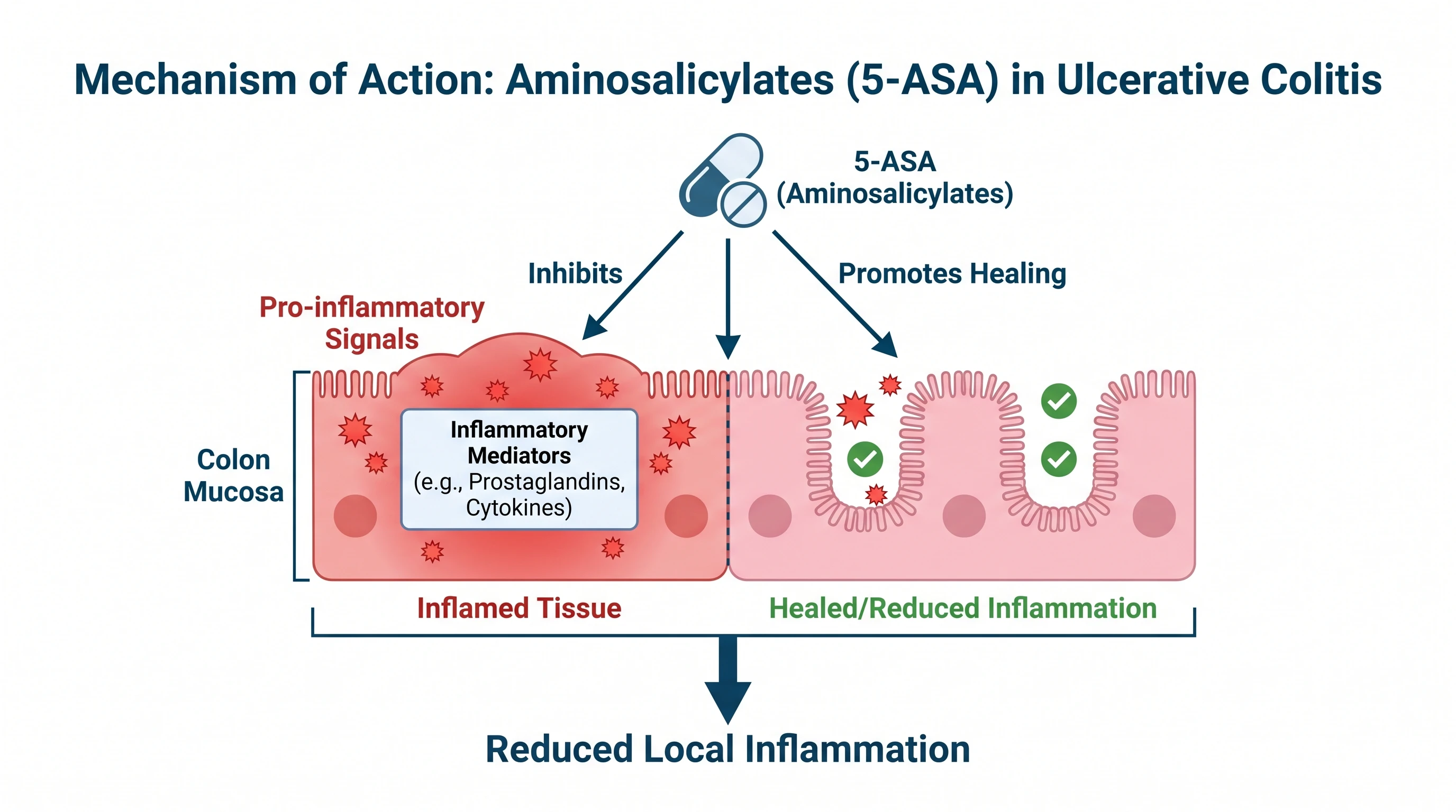
Mesalamine works at the epithelial surface, which is why delivery matters more than dose: azo-bonded prodrugs such as sulfasalazine and balsalazide are cleaved by bacterial azoreductase in the colon, pH-dependent and MMX coatings release distally, and suppositories and enemas cover proctitis and left-sided disease. The mechanism arrows — PPAR-γ activation, NF-κB and eicosanoid suppression — belong inside the epithelial cell. Systemic exposure is a side-effect story, not the mechanism.
Draw gut-selective trafficking at the endothelial wall
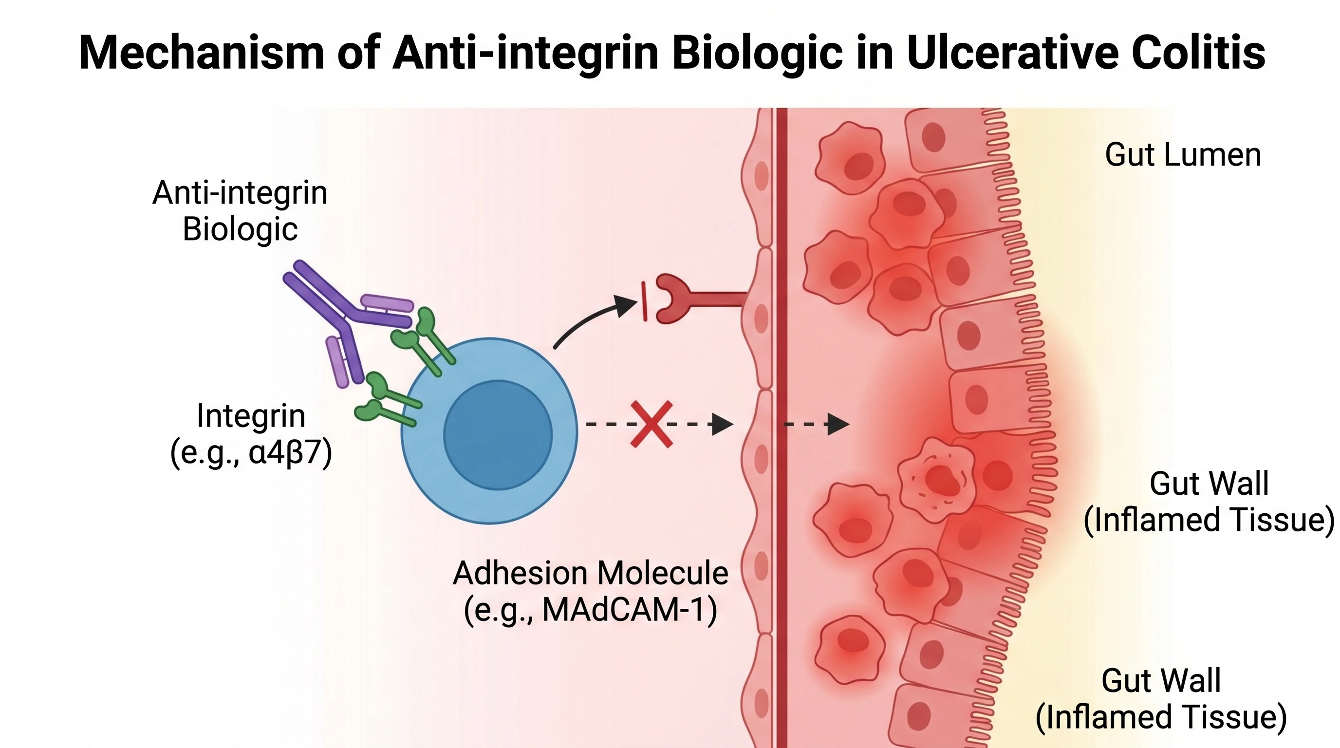
Vedolizumab blocks α4β7 on circulating lymphocytes from binding MAdCAM-1 on intestinal endothelium, so the effect is confined to the gut — the reason it carries a different safety profile from systemic immunosuppression. S1P receptor modulators such as ozanimod and etrasimod act one step earlier, holding lymphocytes inside lymph nodes. Both belong on the vessel, not in the lamina propria, and mixing them up is a common error.
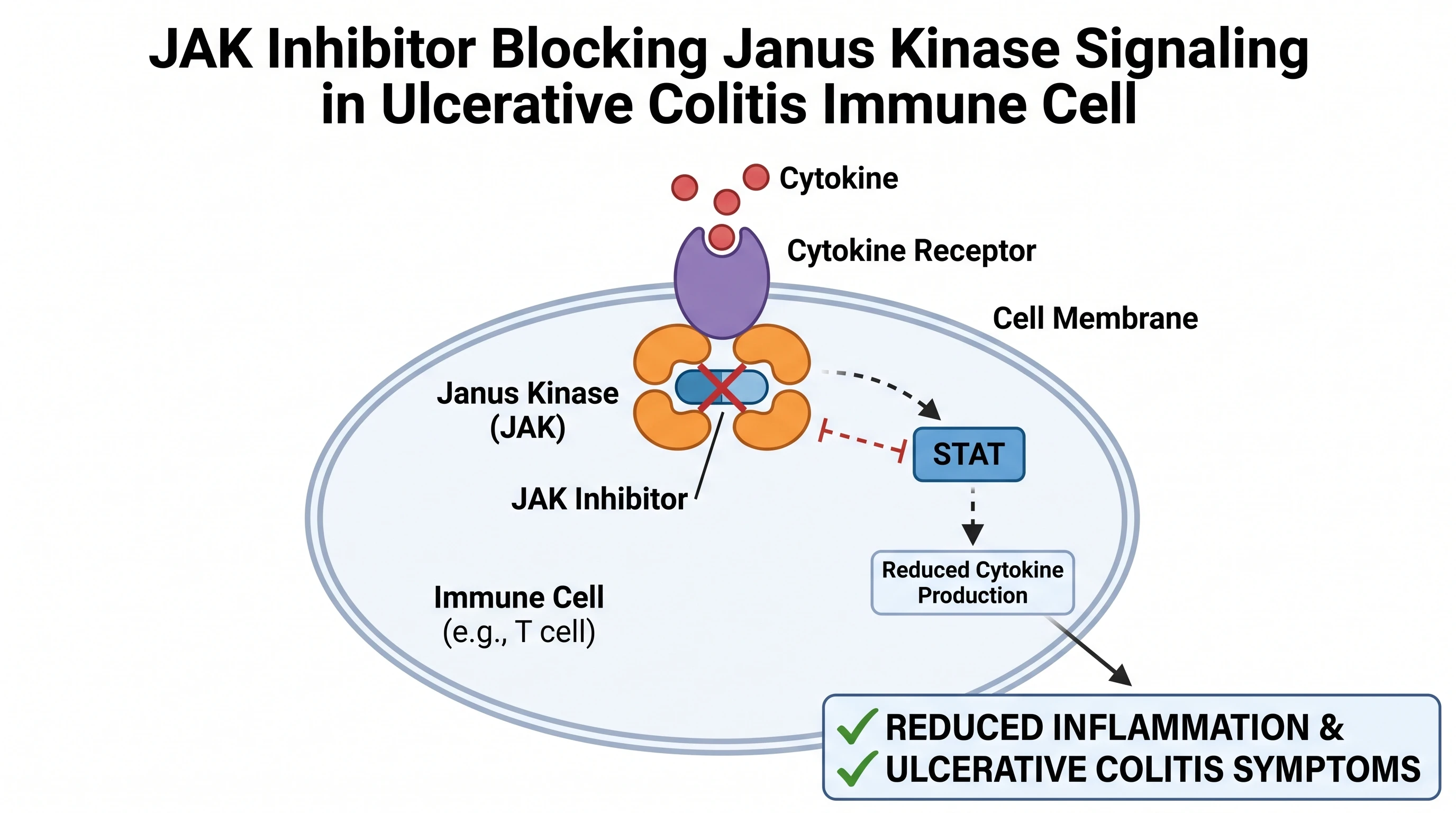
Put the small molecules inside the cell
Tofacitinib and upadacitinib are the only agents in the figure that cross the membrane and act on intracellular signalling: they block JAK–STAT transduction downstream of the common gamma-chain cytokines, IL-6, and interferon-γ, rather than neutralising a single ligand outside the cell. Drawing that distinction — extracellular antibody versus intracellular kinase inhibitor — is what makes the panel worth more than a list of drug names.
Drugs Acting on Various Pathophysiological Pathways in Ulcerative Colitis— templates & examples
How to make Drugs Acting on Various Pathophysiological Pathways in Ulcerative Colitis
Describe your figure
Tell SciFig what to draw in plain language — no design tools required.
Generate with SciFig
Get a clean, publication-ready figure that matches your description in seconds.
Edit & export
Vectorize it into editable SVG, relabel everything, and export for your paper, poster, or slides.
Related searches
- ulcerative colitis medications pathways
- ulcerative colitis drug targets
- ulcerative colitis treatment diagram
- uc drug mechanism of action
- ulcerative colitis biologics
- ibd drug targets
Make figures for
Related pages
Pharmacology Templates
Browse more Pharmacology templates in SciFig's library.
All Templates
Browse every template page in SciFig's library.
Chemotherapeutic Agents Can Classify According to the Mechanism of Action
A publication-ready figure classifying chemotherapeutic agents by mechanism of action and where they act on the cell cycle.
Graphical Abstract Maker
Summarize your study in one panel
Scientific Poster Generator
Design a conference research poster
AI Scientific Image Generator
Generate research figures from text
Conceptual Framework Generator
Map variables and their relationships
Frequently Asked Questions
Common questions about Drugs Acting on Various Pathophysiological Pathways in Ulcerative Colitis.

Ready to publish?
Make your own Drugs Acting on Various Pathophysiological Pathways in Ulcerative Colitis in minutes.
Start for freeFree to start · No credit card required · Built for researchers