
Imaging Flow Cytometer
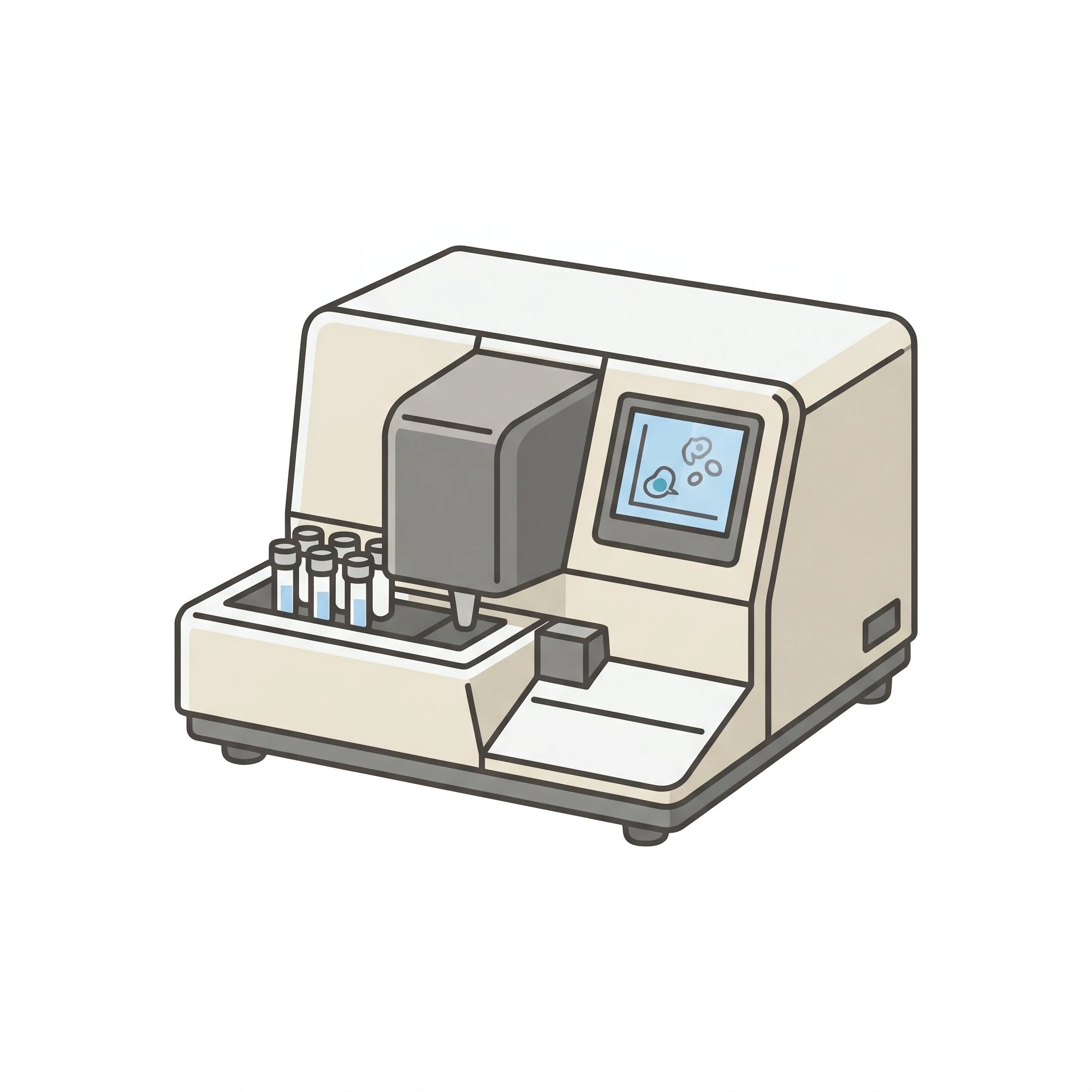
A clean, editable icon of a benchtop imaging flow cytometer — ready for your lab equipment and methods figures.
Updated

Start with up to 150 free credits|No credit card required
Get up to 350 free credits on day one when you join through an invite.
What is Imaging Flow Cytometer?
An imaging flow cytometer is a benchtop instrument that combines flow cytometry with microscopy, capturing an image of every cell as it flows past the optics so researchers measure fluorescence and see cell morphology at once. This icon shows a clean benchtop imaging flow cytometer for equipment and methods figures. With SciFig you describe the instrument in plain language and generate an editable flow cytometer icon you can relabel, recolor, and export as SVG or PNG.
Why the instrument gets its own glyph in a methods figure
- The whole point of the platform is that it is not a conventional cytometer: it records a spatially resolved image of each cell rather than a single integrated intensity, so a generic cytometer silhouette misstates the method.
- Readers need to know from the schematic alone that morphology-derived features (nuclear translocation, internalisation, spot counts, cell–cell conjugates) were available — that expectation is set by the instrument drawn at the top of the pipeline.
- Photographs of benchtop hardware are vendor-specific and date fast; a neutral line drawing survives the next hardware refresh and avoids implying endorsement.
- Methods reproducibility guidance (MIFlowCyt) asks for the instrument, optical configuration, and acquisition settings to be stated; a labeled schematic carries the optical layout compactly alongside that text.
What to include in the instrument or optics diagram
- Sample loading: tube or multi-well plate on an autosampler, feeding a peristaltic or syringe-driven input.
- The flow cell with hydrodynamic focusing — core sample stream sheathed by sheath fluid, narrowing cells into single file. In imaging systems the core is confined in two axes to keep every cell in the focal plane.
- Excitation lasers (typically 405, 488, 561, and 642 nm lines) entering the flow cell orthogonally, plus a brightfield illumination LED on the opposite side and a side-scatter (darkfield) laser.
- Collection optics: a high-NA objective (20×/40×/60×) and a spectral decomposition element — a stacked filter or prism — that fans the signal into parallel channels on one sensor.
- The TDI (time-delay integration) camera, drawn as a sensor whose charge is clocked in step with cell velocity. This is the defining component: it is what allows a moving object to be imaged without motion blur at ~1000 cells/s.
- Channel outputs to label: brightfield, darkfield/SSC, and the fluorescence channels — showing the per-cell image montage as the output makes the imaging modality explicit.
Figures the icon belongs in
- Immunology and cell-biology method panels where the readout is spatial: NF-κB or NFAT nuclear translocation, phagocytosis and internalisation scoring, immunological synapse formation.
- Graphical abstracts for morphology-based assays — apoptosis by nuclear condensation, autophagy puncta counts, micronucleus scoring, DNA-damage foci.
- Rare-event and doublet-discrimination workflows, where per-cell imagery is used to confirm that a gated event is a real cell and not an aggregate or debris.
- Equipment and core-facility figures: instrument inventory slides, service catalogues, and grant facility descriptions.
- Pipeline diagrams that carry through to downstream analysis — feature extraction, masks, and classifier training on the per-cell image set.
Imaging Flow Cytometer— templates & examples
How to make Imaging Flow Cytometer
Describe your figure
Tell SciFig what to draw in plain language — no design tools required.
Generate with SciFig
Get a clean, publication-ready figure that matches your description in seconds.
Edit & export
Vectorize it into editable SVG, relabel everything, and export for your paper, poster, or slides.
Related searches
- imaging flow cytometer icon
- flow cytometer icon
- flow cytometry instrument icon
- cytometer machine icon
- lab instrument icon
- imaging flow cytometry
Make figures for
Related pages
Lab Equipment Icons
Browse more Lab Equipment icons in SciFig's library.
All Icons
Browse every icon page in SciFig's library.
Coplin Jar with Blood Smear Slides
A clean, editable icon of a Coplin staining jar holding blood smear slides — ready for your histology and microscopy figures.
Magnetic Cell Separator
A clean, editable magnetic cell separator for immunology and cell-isolation figures — a column in a magnet retaining magnetically labeled cells, ready to relabel.
Cubitainer Container 5 Gallon
A clean, editable icon of a 5-gallon cubitainer — the collapsible cube-shaped container used for lab liquids and samples.
AI Scientific Image Generator
Generate research figures from text
Graphical Abstract Maker
Summarize your study in one panel
Text to Diagram Generator
Turn text into a diagram
Scientific Poster Generator
Design a conference research poster
Frequently Asked Questions
Common questions about Imaging Flow Cytometer.

Ready to publish?
Make your own Imaging Flow Cytometer in minutes.
Start for freeFree to start · No credit card required · Built for researchers