
Magnetic Cell Separator
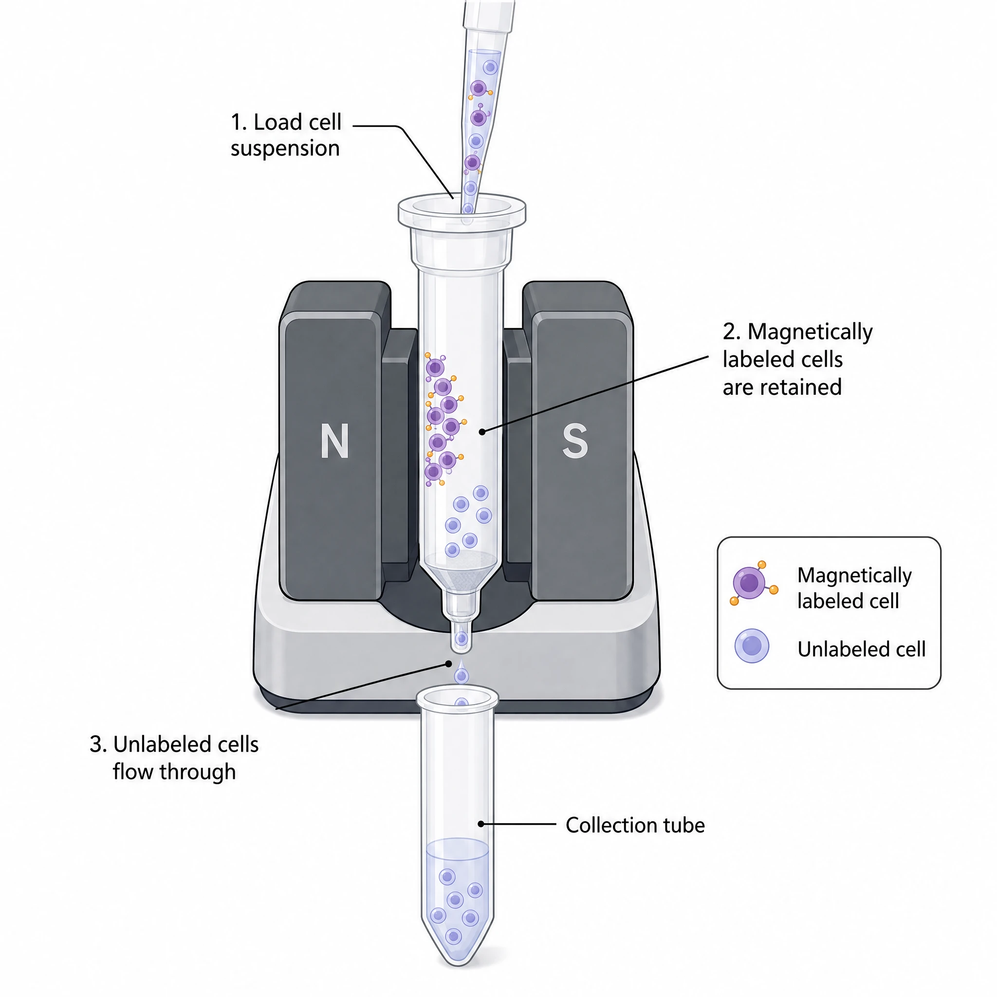
A clean, editable magnetic cell separator for immunology and cell-isolation figures — a column in a magnet retaining magnetically labeled cells, ready to relabel.
Updated

Start with up to 150 free credits|No credit card required
Get up to 350 free credits on day one when you join through an invite.
What is Magnetic Cell Separator?
A magnetic cell separator is a benchtop instrument that isolates target cells by passing a cell suspension through a column held in a strong magnet. Cells tagged with magnetic beads are retained in the column while unlabeled cells flow through, enabling positive or negative selection. It is a common icon in immunology, stem-cell, and cell-therapy figures. With SciFig you describe the separator in plain language and generate a clean, editable magnetic cell separator, ready to relabel and export.
Why the separation step is drawn out
- The direction of the enrichment is the claim. Positive selection (target retained on the column, labeled) and negative selection (target passes through, untouched) yield functionally different cells, and only a diagram makes clear which one was done.
- Bead retention on positively selected cells can occlude the epitope and even signal through it, so a figure that does not indicate the selection mode leaves a real confound unstated.
- It anchors purity claims: a schematic that shows starting material → labeling → column → flow-through and eluate gives the reader a place to hang the pre/post purity percentages from the cytometry panel next to it.
- It is a column-and-magnet method, not a sorter. Conflating it with FACS in a workflow figure misstates the throughput (10⁸–10⁹ cells in minutes, bulk, no per-cell decision) and the level of resolution achievable.
Parts of the schematic
- The permanent magnet assembly and the column seated in its gap — the ferromagnetic matrix or spheres inside the column are what amplify the field into steep local gradients, and drawing that matrix is what explains why the cells stick.
- The magnetic label itself: antibody-conjugated superparamagnetic beads. Size is a real distinction to encode — nanoscale beads (~50 nm) are designed to be left on the cell, while larger micron-scale beads generally require a release or detachment step.
- Direct versus indirect labeling: an antibody-bead conjugate binding the target antigen directly, versus a primary antibody (often biotinylated) followed by an anti-biotin or secondary bead.
- The two output fractions, drawn and named without ambiguity: the flow-through (unlabelled cells) and the eluate (labeled cells, released only after the column is taken out of the magnet and flushed with the plunger).
- The selection mode, shown as two parallel branches: positive selection labels the target; negative selection labels everything else with a lineage-depletion cocktail so the target is never touched.
- Optional but informative: buffer composition cues (degassed, EDTA-containing, cold) and a pre-filter, since clumps and DNA from dead cells are the standard cause of column clogging.
Figures it belongs in
- Immunology methods: PBMC isolation followed by CD4⁺, CD8⁺, CD14⁺, or CD19⁺ enrichment, and untouched naive T cell isolation by depletion cocktail.
- Stem-cell and haematology workflows — CD34⁺ progenitor enrichment from apheresis or marrow, and lineage-negative depletion prior to sorting.
- Cell-therapy manufacturing schematics: T cell selection at the front of a CAR-T process, where enrichment is a GMP unit operation, not a bench prep.
- Tumour dissociation pipelines: dead-cell removal, red-cell depletion, and CD45 depletion to enrich tumour cells before single-cell sequencing.
- Two-step protocol figures where magnetic pre-enrichment feeds a downstream sorter — a common design that raises rare-event yield without the sorter's time cost.
- Protocol SOPs and training slides where the column-in-magnet / column-out-of-magnet distinction is the entire operational point.
Magnetic Cell Separator— templates & examples
How to make Magnetic Cell Separator
Describe your figure
Tell SciFig what to draw in plain language — no design tools required.
Generate with SciFig
Get a clean, publication-ready figure that matches your description in seconds.
Edit & export
Vectorize it into editable SVG, relabel everything, and export for your paper, poster, or slides.
Related searches
- magnetic cell separator
- magnetic cell separation
- macs cell separation column
- magnetic bead cell sorting
- cell isolation magnet
- magnetic separation column icon
- immunomagnetic separation diagram
- cell sorting equipment icon
- immunomagnetic column separator
- macs separator icon
Make figures for
Related pages
Lab Equipment Icons
Browse more Lab Equipment icons in SciFig's library.
All Icons
Browse every icon page in SciFig's library.
Coplin Jar with Blood Smear Slides
A clean, editable icon of a Coplin staining jar holding blood smear slides — ready for your histology and microscopy figures.
Cubitainer Container 5 Gallon
A clean, editable icon of a 5-gallon cubitainer — the collapsible cube-shaped container used for lab liquids and samples.
Imaging Flow Cytometer
A clean, editable icon of a benchtop imaging flow cytometer — ready for your lab equipment and methods figures.
AI Scientific Image Generator
Generate research figures from text
Graphical Abstract Maker
Summarize your study in one panel
Text to Diagram Generator
Turn text into a diagram
Scientific Poster Generator
Design a conference research poster
Frequently Asked Questions
Common questions about Magnetic Cell Separator.

Ready to publish?
Make your own Magnetic Cell Separator in minutes.
Start for freeFree to start · No credit card required · Built for researchers