Mouse Brain Anatomy: Dorsal and Ventral Views
A labeled mouse brain anatomy figure showing dorsal and ventral views for neuroscience papers, posters, and slides — relabel and export in any format.
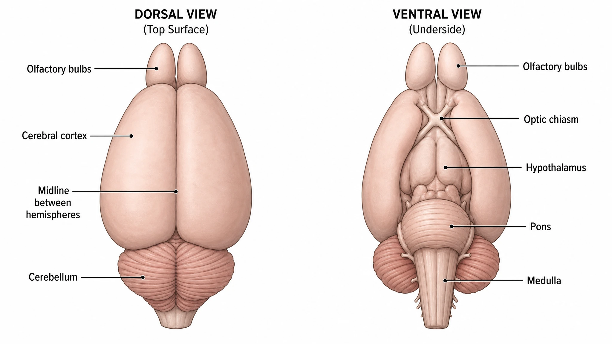
Figure prompt
Core Subject (e.g., Cas9 protein cutting DNA)
Action / Details (e.g., Double strand break, detailed molecular view)
Start with 200 free credits|No credit card required
Get up to 400 free credits on day one when you join through an invite.
What is Mouse Brain Anatomy: Dorsal and Ventral Views?
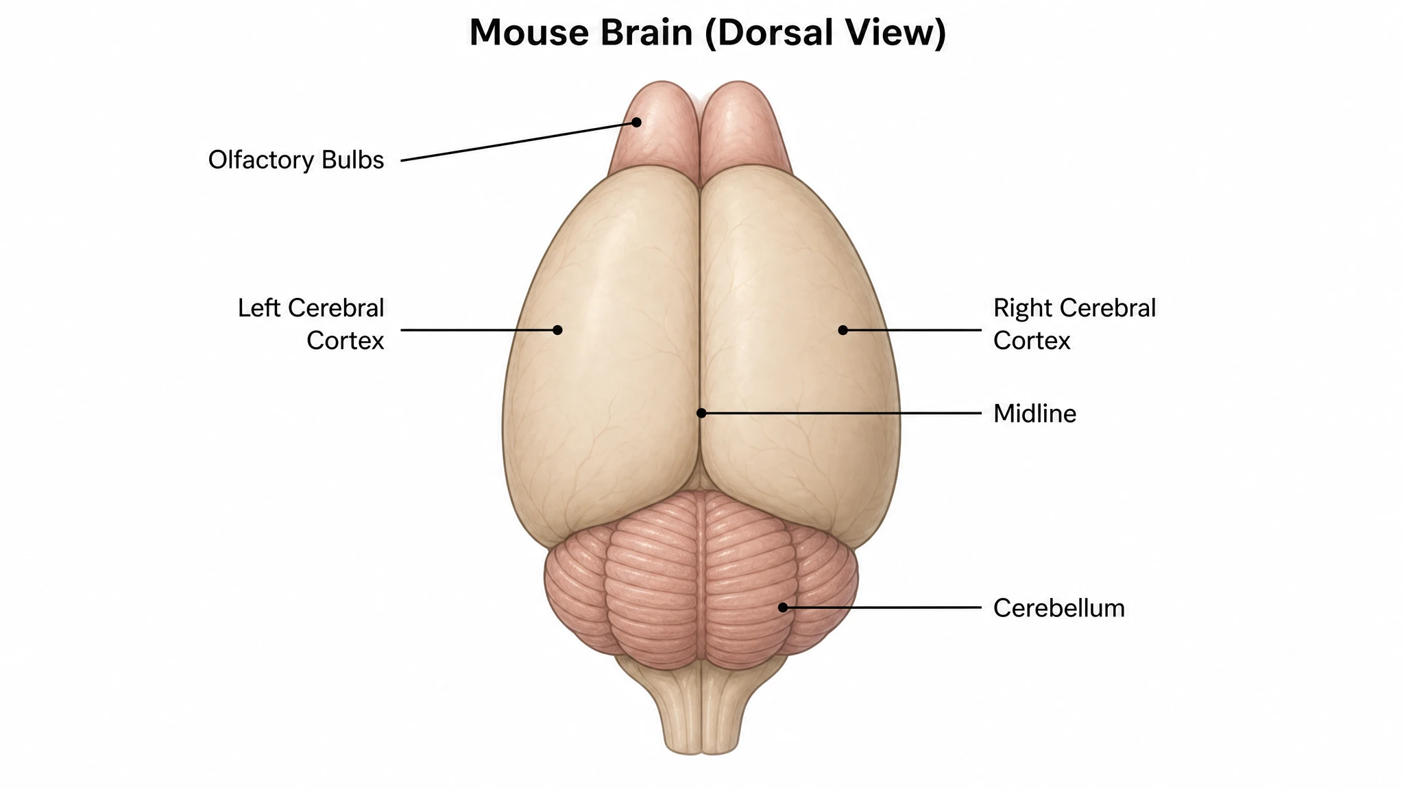
A mouse brain anatomy diagram is a labeled illustration of the mouse (Mus musculus) brain shown from the dorsal and ventral surfaces. The dorsal view shows the olfactory bulbs, the cerebral cortex, the midline between the two hemispheres, the cerebellum, and the brainstem; the ventral view shows the olfactory bulbs, optic chiasm, hypothalamus, pons, and medulla. With SciFig you describe the views you need and generate a clean, publication-ready mouse brain figure you can relabel and export.
Why Researchers Draw This Figure
- Surface views orient every downstream figure in a paper — injection sites, implant placements and sectioning planes are all reported relative to them.
- The two surfaces expose disjoint sets of structures: the visual and hypothalamic landmarks are only on the ventral face, the collicular and cerebellar landmarks only on the dorsal, so one view alone is never sufficient.
- Rodent and human anatomy differ in ways that matter for translational claims — a smooth cortex, a proportionally enormous olfactory bulb, an exposed midbrain roof — and the drawing states those differences instead of leaving them implied.
- Photographs of a dissected specimen are variable, glare-prone and hard to label cleanly; a schematic is reproducible across every figure in a lab.
- Stereotaxic surgery is described relative to skull landmarks, and a diagram that includes bregma and lambda ties the coordinate frame to the anatomy it targets.
- Atlas plates from Paxinos and Franklin or the Allen reference are copyrighted; an original labeled schematic sidesteps the permissions problem entirely.
Structures and Landmarks to Label
- Olfactory bulbs and tracts — visible from both surfaces and disproportionately large in this species, occupying a far greater fraction of the forebrain than in primates.
- Cerebral cortex — smooth and gyrus-free, divided by the interhemispheric fissure, with functional areas indicated by position rather than by sulcal boundaries.
- Superior and inferior colliculi — the midbrain roof, exposed dorsally between the caudal cortex and the cerebellum.
- Cerebellum — vermis flanked by hemispheres and paraflocculi, with the transverse folia and the primary fissure as the reliable dorsal landmarks.
- Optic chiasm and hypothalamic floor — the ventral fixed points, together with the tuber cinereum, median eminence, pituitary stalk and mammillary bodies.
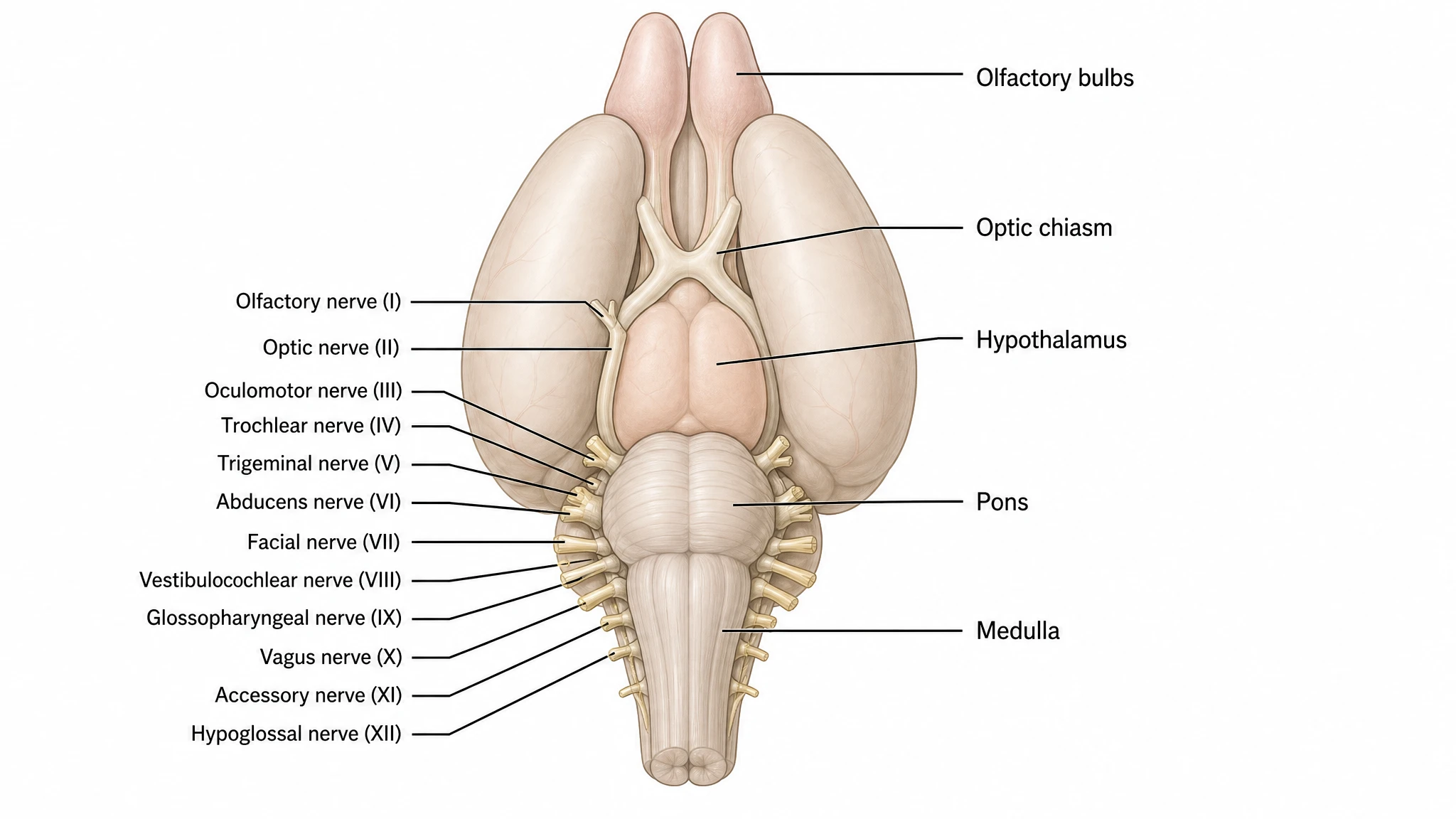
- Brainstem — cerebral peduncles, the transverse pontine fibers, the medullary pyramids, and the emerging cranial nerve roots including the prominent trigeminal.
- Stereotaxic frame — bregma and lambda on the skull with anteroposterior, mediolateral and dorsoventral axes; in an adult C57BL/6 the bregma–lambda distance is roughly 4.2 mm, and stating strain and age is what makes published coordinates usable.
Where This Figure Is Used
- Methods figures for stereotaxic viral injection, showing target coordinates and the intended spread of an AAV or tracer.
- Optogenetic and fiber photometry papers, where implant trajectory and fiber tip position must be shown relative to surface landmarks.
- Dissection and microdissection protocols that specify which structures are removed and in what order, for regional transcriptomics or proteomics.
- Perfusion, embedding and sectioning documentation — defining coronal, sagittal or horizontal planes against a surface reference so section figures can be located.
- Lesion, electrode and cannula placement verification, where a schematic plus histology is the standard evidence pairing.
- Teaching and comparative neuroanatomy, contrasting rodent organization with human or non-human primate organization on matched views.
What This Template Gives You
Dorsal view with the lissencephalic cortex labeled
The view from above shows the olfactory bulbs projecting well forward of the cerebrum, the smooth unfissured cortical surface split by the interhemispheric fissure, the superior and inferior colliculi exposed between cortex and cerebellum, and the foliated cerebellar vermis and hemispheres. That exposed midbrain roof is a real anatomical difference from primates, where the occipital cortex covers it, and the figure labels it rather than hiding it.
Ventral view from olfactory tract to medulla
The inferior surface carries the landmarks that orient any perfusion or dissection: olfactory tracts, the optic chiasm, the tuber cinereum and median eminence with the pituitary stalk, the mammillary bodies, the cerebral peduncles, the transverse fibers of the pons, and the pyramids of the medulla. Cranial nerve roots — the large trigeminal in particular — are marked where they emerge, since they are the fastest way to confirm orientation on a fresh specimen.
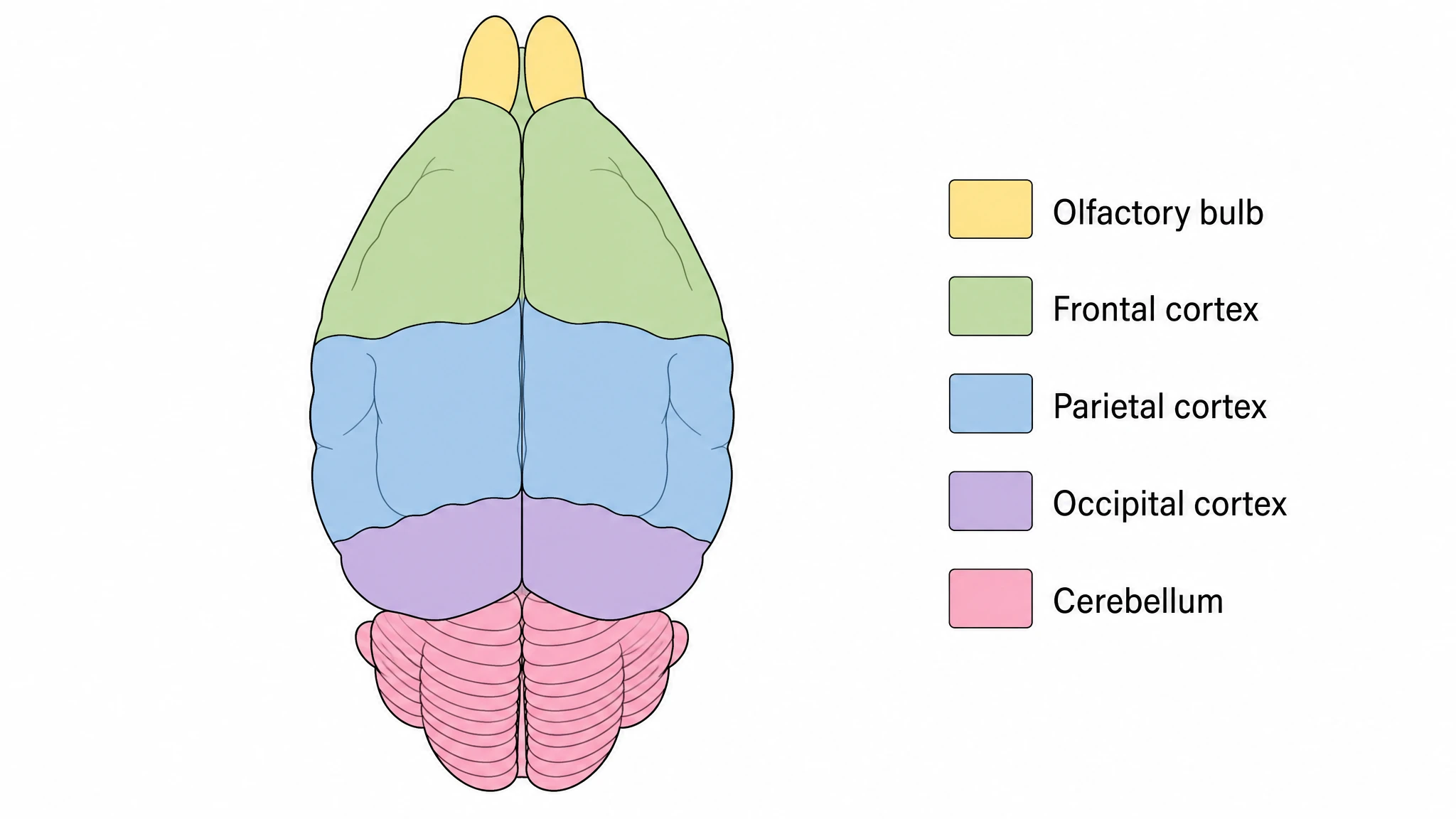
Color-coded major divisions across both surfaces
A single palette maps telencephalon, diencephalon, midbrain, cerebellum and brainstem consistently on the dorsal and ventral panels, so a region keeps its color wherever it appears. This is what makes the two views readable as one specimen instead of two unrelated drawings, and the same key can be carried through to section figures later in a paper for visual continuity.
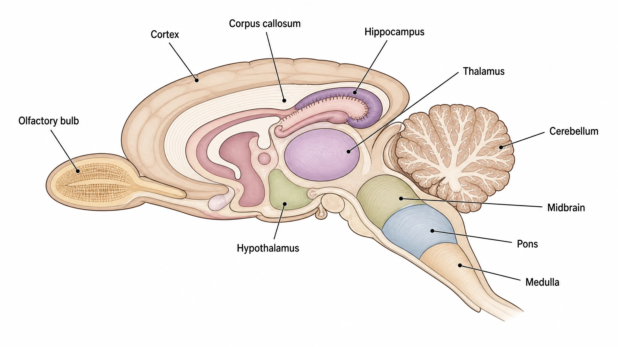
Midsagittal section for the internal reference
The cut in the midline exposes what neither surface view can show: corpus callosum, the ventricular system, thalamus and hypothalamus, the cerebral aqueduct, and the cerebellar arbor vitae. Placed next to the surface panels it gives depth context for stereotaxic targeting, where dorsoventral coordinates are meaningless without knowing what lies between the skull surface and the intended structure.
Mouse Brain Anatomy: Dorsal and Ventral Views— templates & examples
How to make Mouse Brain Anatomy: Dorsal and Ventral Views
Describe your figure
Tell SciFig what to draw in plain language — no design tools required.
Generate with SciFig
Get a clean, publication-ready figure that matches your description in seconds.
Edit & export
Vectorize it into editable SVG, relabel everything, and export for your paper, poster, or slides.
Related searches
- mouse brain
- mouse brain anatomy
- mouse brain dorsal view
- mouse brain ventral view
- mouse brain diagram labeled
- mouse brain regions
- mouse brain atlas
- mus musculus brain anatomy
- mouse brain illustration
- labeled mouse brain figure
Make figures for
Frequently Asked Questions
Common questions about Mouse Brain Anatomy: Dorsal and Ventral Views.

Ready to publish?
Make your own Mouse Brain Anatomy: Dorsal and Ventral Views in minutes.
Start for freeFree to start · No credit card required · Built for researchers