
Different Types of Nanoparticles
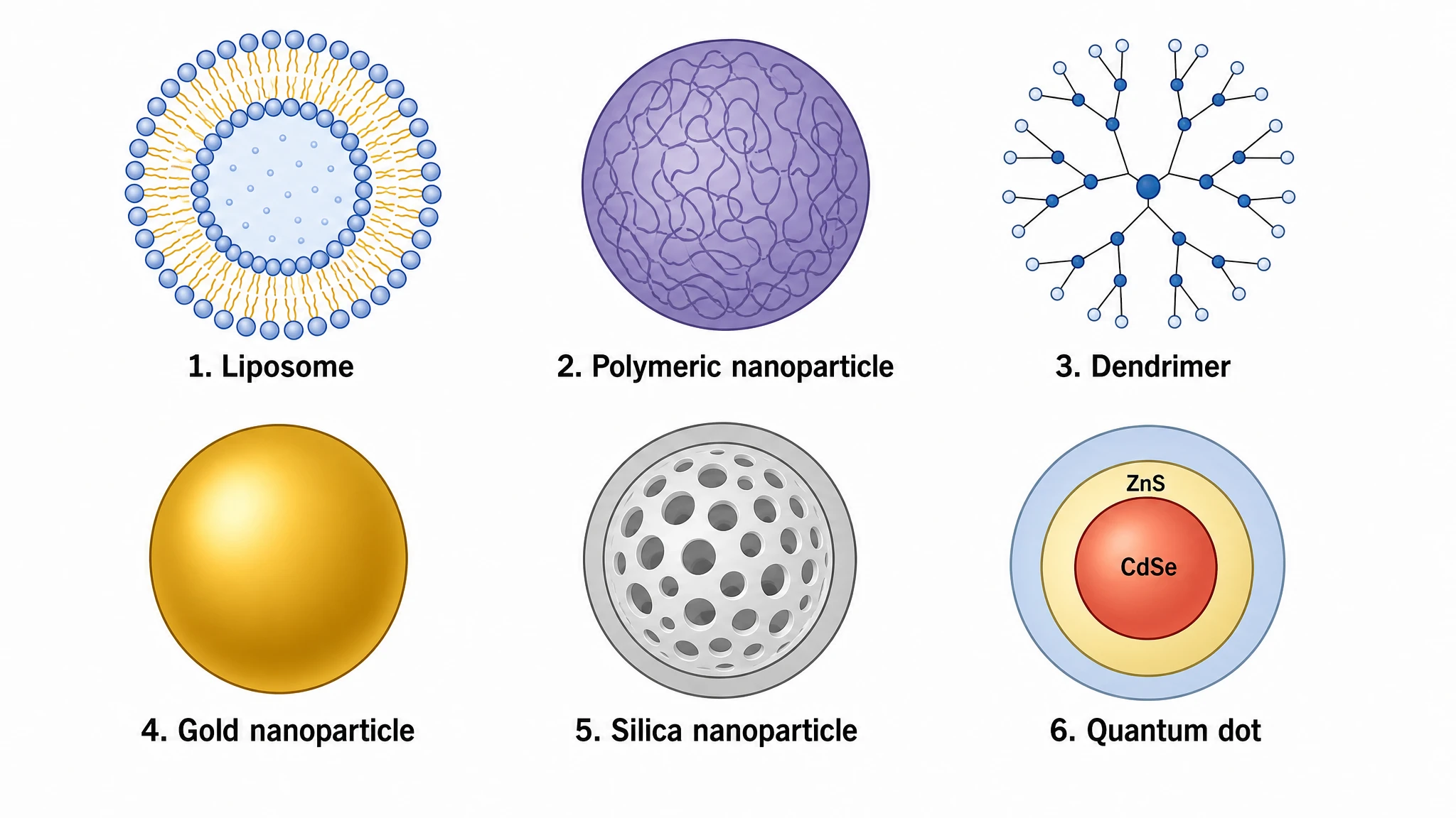
An editable comparison figure showing the main types of nanoparticles — liposomes, polymeric, gold, quantum dots, and dendrimers — side by side with their distinct structures.
Updated

Start with up to 150 free credits|No credit card required
Get up to 350 free credits on day one when you join through an invite.
What is Different Types of Nanoparticles?
A types-of-nanoparticles figure compares the main classes of engineered nanoparticles side by side. It typically shows lipid-based particles such as liposomes, polymeric nanoparticles and dendrimers, inorganic particles such as gold nanoparticles and silica, and semiconductor quantum dots, highlighting each particle's structure, core, and surface. With SciFig you describe the nanoparticle classes in plain language and generate a clean, editable comparison figure you can relabel and export.
Why Researchers Draw This Figure
- A side-by-side panel is the fastest way to justify a carrier choice in an introduction — it shows what was considered and rejected, not just what was used.
- Drawn at a shared scale, the classes reveal a two-order-of-magnitude spread (a 3 nm dot next to a 200 nm vesicle), which a table of diameters never communicates.
- Every class shares the same three-part anatomy — core, shell or matrix, surface layer — and the figure makes that common grammar visible instead of leaving each panel to invent its own.
- Function follows the core, but fate in vivo follows the surface: separating those two layers graphically prevents the common error of attributing circulation time to the material rather than the coating.
- Grant reviewers and thesis committees expect a landscape figure before a mechanism figure; this one carries that job.
- Cartoons copied from vendor slide decks are usually structurally wrong (bilayers drawn as single lines, dendrimers drawn as spheres) and are not licensable — an original schematic avoids both problems.
Classes and Features to Label
- Liposomes and lipid nanoparticles — phospholipid bilayer vesicles (50–200 nm) or ionizable-lipid LNPs; the clinical mainstay for doxorubicin delivery and for mRNA payloads.
- Polymeric nanoparticles and micelles — PLGA or PCL matrices and amphiphilic block copolymers that self-assemble above a critical micelle concentration, with cargo release governed by matrix degradation rather than diffusion.
- Dendrimers — covalently branched, monodisperse architectures (PAMAM G3–G5, roughly 3–7 nm) with a defined number of terminal groups; the only class with an exact molecular weight rather than a distribution.
- Gold nanoparticles — spheres, rods, stars and shells (5–100 nm), thiol-functionalized, exploited for plasmonic heating, scattering contrast and label-free sensing.
- Iron oxide (SPIONs) — superparamagnetic magnetite or maghemite cores under ~20 nm, used as T2 MRI contrast and for magnetic hyperthermia and cell sorting.
- Quantum dots and carbon nanomaterials — semiconductor cores with size-tunable emission; carbon nanotubes and graphene oxide with high aspect ratio and strong NIR absorbance.
- Surface layer and colloidal descriptors — PEGylation density, targeting ligands, zeta potential (a magnitude above roughly ±30 mV predicts colloidal stability), hydrodynamic diameter versus core diameter, and the protein corona that forms on contact with serum.
Where This Figure Is Used
- Drug delivery papers that must place a chosen carrier in context, including passive tumor accumulation via the EPR effect and its now well-documented limits in human tumors.
- Vaccine and nucleic-acid delivery work, where ionizable lipid formulations must be contrasted with polymeric and dendritic alternatives for endosomal escape.
- Imaging and contrast-agent studies pairing magnetic cores for MRI with fluorescent or plasmonic cores for optical readout.
- Photothermal and photodynamic therapy, where absorbance in the near-infrared tissue window determines whether a core is usable at all.
- Biosensing and diagnostics: lateral-flow assays, plasmonic colorimetric readouts, and semiconductor labels for multiplexed detection.
- Nanotoxicology and regulatory dossiers, where size, shape, surface charge and biopersistence must be reported per class rather than as a single average.
What This Template Gives You
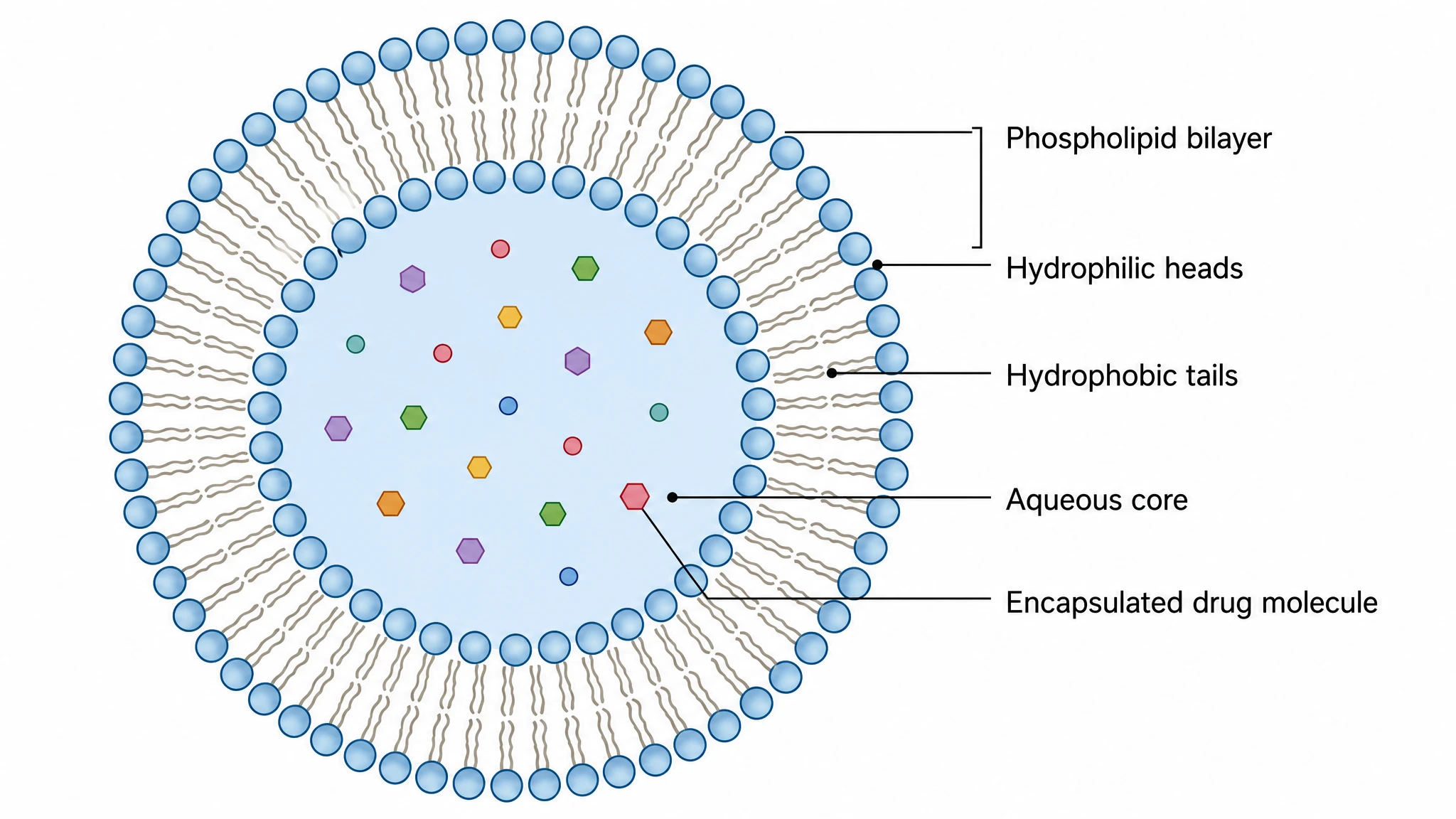
Liposome drawn with a resolved bilayer and core
The lipid vesicle is rendered as a genuine phospholipid bilayer around an aqueous lumen, typically 50–200 nm across, with the two loading compartments made visible: hydrophilic cargo in the core, lipophilic cargo intercalated between the acyl chains. A grafted PEG corona is shown separately from the bilayer, because stealth behavior and circulation half-life come from that brush layer rather than from the lipids themselves.
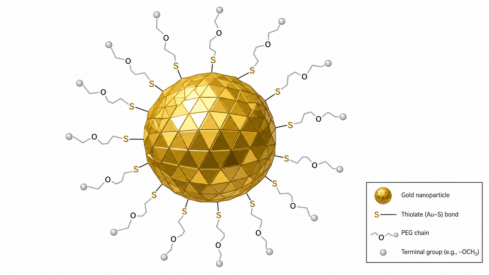
Gold particle with surface chemistry made explicit
A metallic core of roughly 5–100 nm is shown with the thiol-anchored ligand shell that defines its colloidal behavior. Callouts mark the surface plasmon resonance near 520 nm for small spheres, and the red-shift into the near-infrared window when the geometry becomes a rod or a shell — the property that carries photothermal and photoacoustic applications. Citrate, PEG-thiol and antibody conjugates are drawn as distinct outer layers.
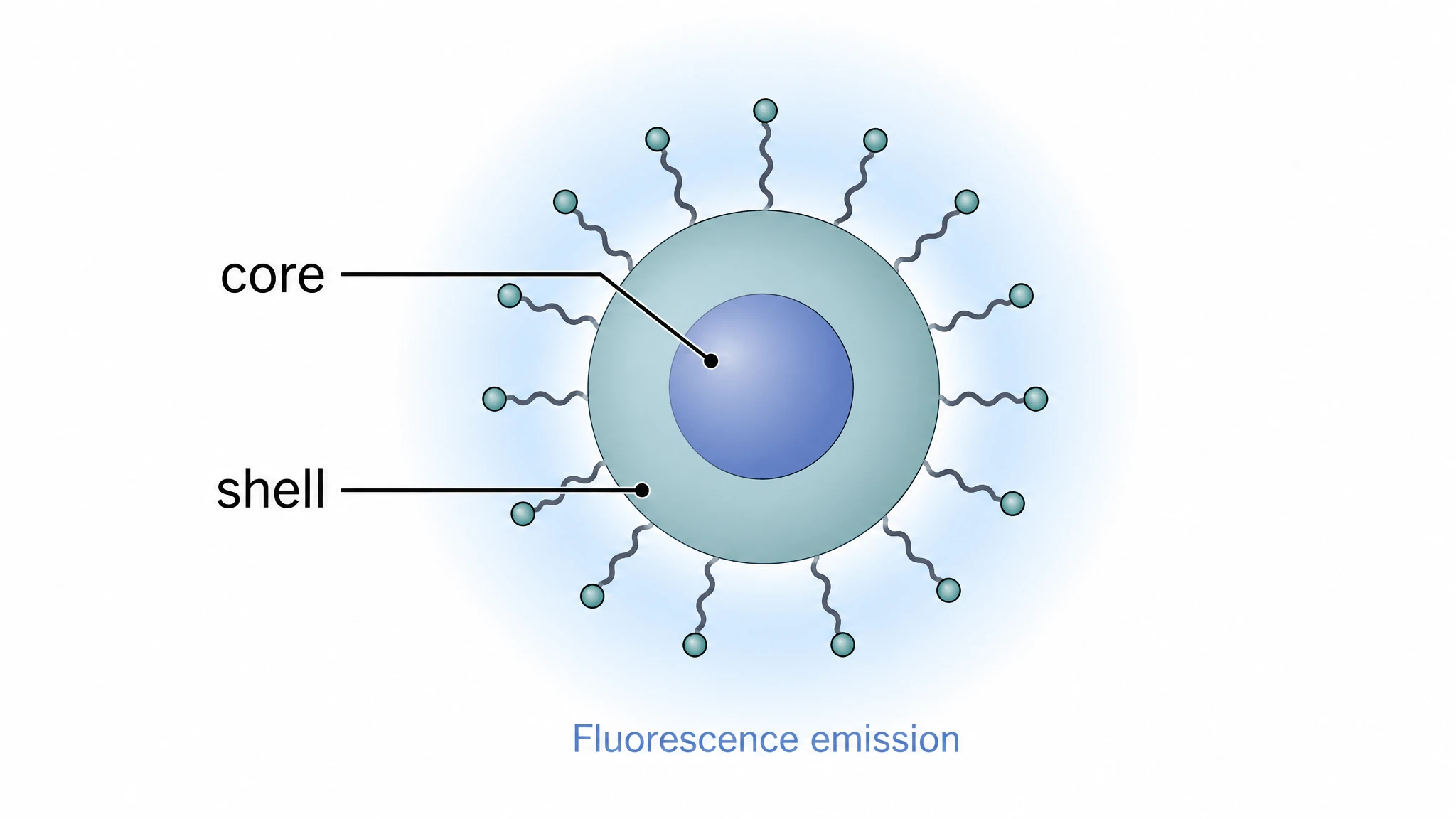
Quantum dot as a core–shell semiconductor
The semiconductor is drawn as a 2–10 nm crystalline core with a wider-bandgap shell, usually CdSe/ZnS or an InP alternative, plus a solubilizing ligand layer. An emission strip shows size-tunable fluorescence: quantum confinement means a smaller core emits blue and a larger one emits red from the same material, which is the single fact that most distinguishes these from any other label in the figure.
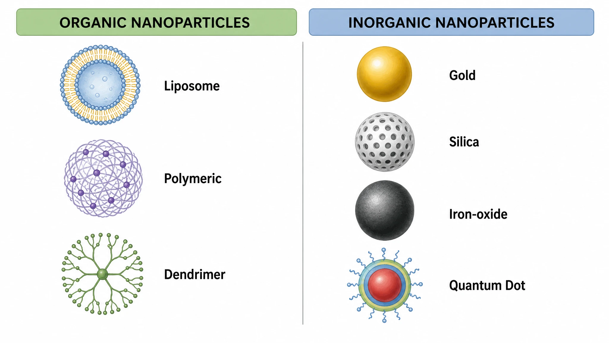
Organic versus inorganic classification axis
The final panel arranges the classes along the split that actually governs experimental choice. Organic carriers — lipid, polymeric, dendritic — are biodegradable, carry high payloads, and are cleared by hepatic and renal routes. Inorganic cores — metallic, oxide, semiconductor — bring optical, magnetic or catalytic function that no soft carrier provides, at the cost of persistence and a longer regulatory path. Size and surface charge are annotated on both sides.
Different Types of Nanoparticles— templates & examples
How to make Different Types of Nanoparticles
Describe your figure
Tell SciFig what to draw in plain language — no design tools required.
Generate with SciFig
Get a clean, publication-ready figure that matches your description in seconds.
Edit & export
Vectorize it into editable SVG, relabel everything, and export for your paper, poster, or slides.
Related searches
- types of nanoparticles
- types of nanomolecules
- nanoparticle classification
- kinds of nanoparticles
- organic and inorganic nanoparticles
- liposome nanoparticle
- polymeric nanoparticle
- gold nanoparticle
- quantum dot nanoparticle
- nanoparticle types diagram
Make figures for
Related pages
Nanotechnology Templates
Browse more Nanotechnology templates in SciFig's library.
All Templates
Browse every template page in SciFig's library.
Graphical Abstract Maker
Summarize your study in one panel
Scientific Poster Generator
Design a conference research poster
AI Scientific Image Generator
Generate research figures from text
Conceptual Framework Generator
Map variables and their relationships
Frequently Asked Questions
Common questions about Different Types of Nanoparticles.

Ready to publish?
Make your own Different Types of Nanoparticles in minutes.
Start for freeFree to start · No credit card required · Built for researchers