
NLRP3 Inflammasome Pathway
A clear diagram of the NLRP3 inflammasome pathway, from two-signal priming and activation through NLRP3-ASC-caspase-1 assembly to IL-1 beta and IL-18 release.
Updated

Start with up to 150 free credits|No credit card required
Get up to 350 free credits on day one when you join through an invite.
What is NLRP3 Inflammasome Pathway?
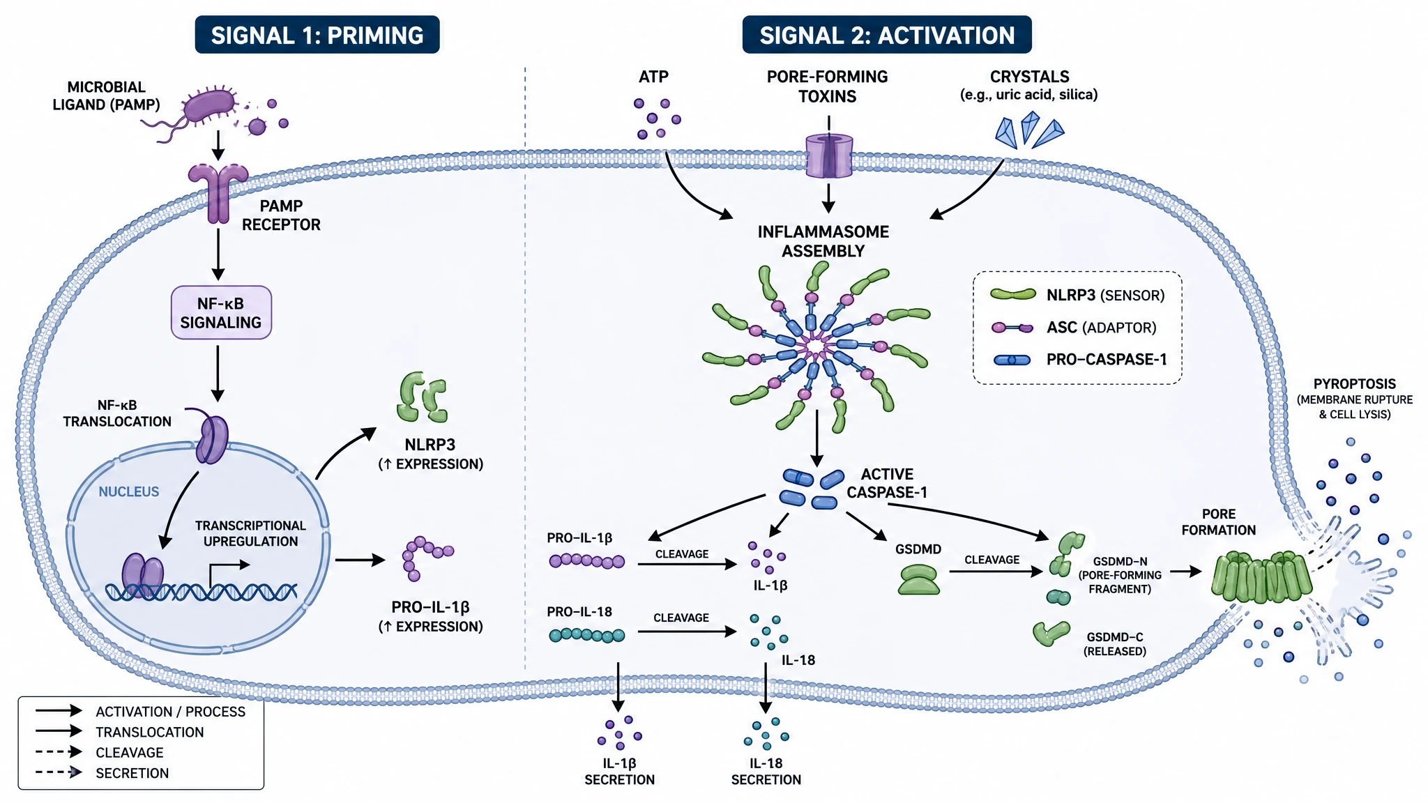
The NLRP3 inflammasome pathway is an innate-immune signaling pathway that drives inflammation after infection and stress. It works in two signals: a priming signal (often via NF-kB) raises NLRP3 and pro-IL-1 beta, and an activation signal triggers assembly of the inflammasome from NLRP3, the ASC adaptor, and pro-caspase-1. Active caspase-1 then cleaves pro-IL-1 beta and pro-IL-18 into mature cytokines and cleaves gasdermin D to drive pyroptosis. With SciFig you build a labeled diagram to export.
Why this signalling cascade is worth diagramming carefully
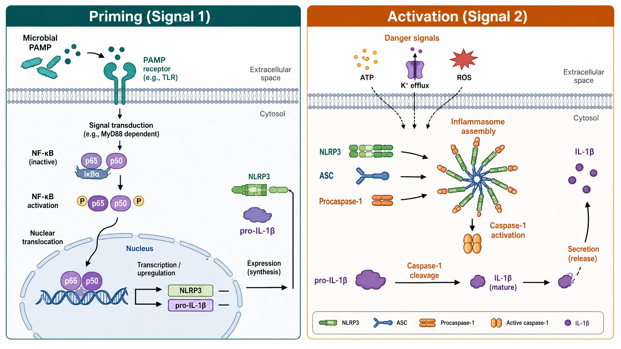
- The two-signal requirement is the most frequently misdrawn part of innate-immune figures. LPS alone primes but does not activate; ATP alone does nothing in an unprimed cell. Collapsing the two into one arrow deletes the mechanism.
- It is a convergence point. Crystals, pore-forming toxins, extracellular ATP and mitochondrial damage all funnel into one sensor, which is exactly why it sits at the center of sterile inflammation.
- Caspase-1 has two outputs — cytokine maturation and gasdermin cleavage — and the figure has to branch there, or it misrepresents pyroptosis as an incidental side effect rather than a parallel arm.
- Release is unconventional and pore-dependent, which changes how secretion inhibitors, supernatant ELISAs and LDH-release readouts should be interpreted.
- It is clinically anchored: gain-of-function mutations in the sensor cause cryopyrin-associated periodic syndromes, and IL-1 blockade with anakinra or canakinumab treats them.
- Small-molecule inhibitors such as MCC950 act at the NACHT domain, so only a domain-level drawing can support a claim about where a compound intervenes.
Components and steps to label
- Signal 1 (priming) — TLR4/MyD88 or TNF receptor signalling to NF-kB, upregulating the sensor and pro-IL-1 beta; plus a rapid non-transcriptional licensing step involving deubiquitination of the sensor.
- Signal 2 (activation triggers) — extracellular ATP acting through P2X7 and the bacterial ionophore nigericin, both driving potassium efflux; particulates including monosodium urate, silica, asbestos, cholesterol crystals and amyloid beta, which rupture lysosomes and release cathepsin B; and mitochondrial ROS with oxidised mtDNA.
- Potassium efflux — the common proximal event shared by most triggers. Raising extracellular potassium blocks activation, which is the standard control experiment and belongs in any mechanistic figure.
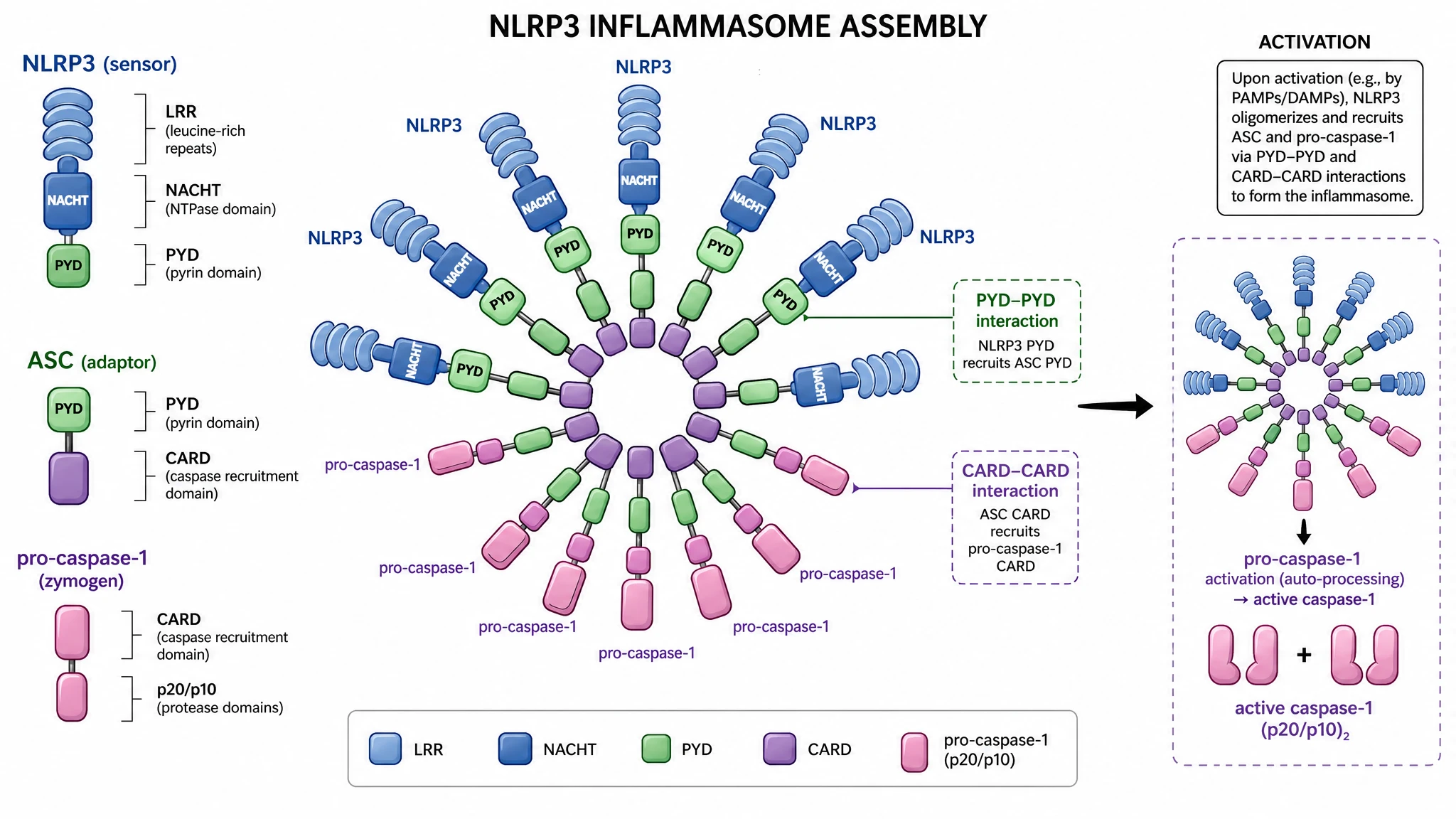
- The sensor — PYD, NACHT (NTPase) and LRR domains, with NEK7 as the licensing partner for oligomerisation and the binding site for MCC950-class inhibitors.
- ASC adaptor — a PYD-CARD bridging protein that nucleates helical filaments and condenses into a single perinuclear speck, widely used as a microscopy readout of activation.
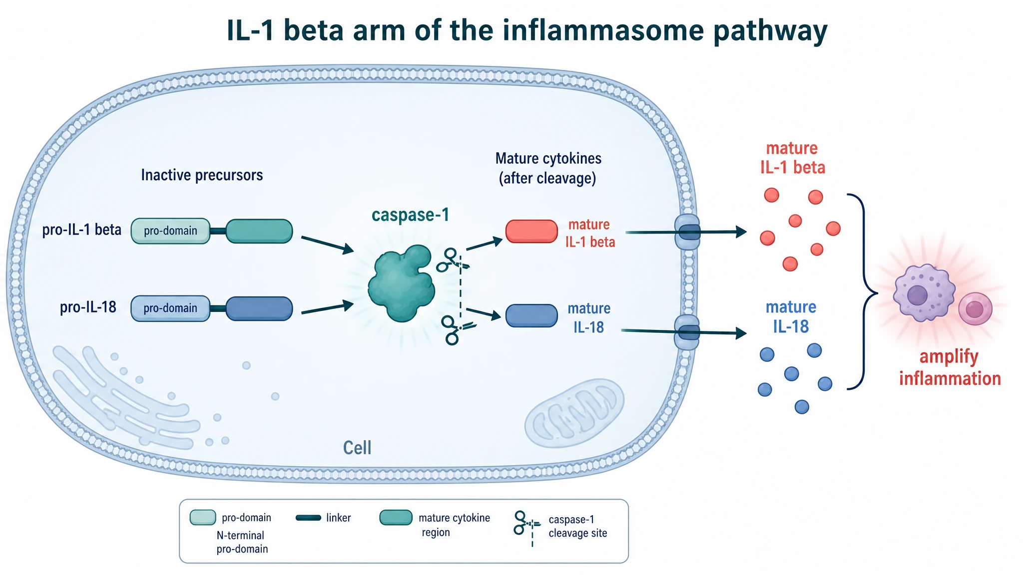
- Caspase-1 — recruited as an inactive zymogen, autoprocessed to p20/p10; its substrates are pro-IL-1 beta, pro-IL-18 and gasdermin D.
- Outputs — mature IL-1 beta and IL-18 exported through gasdermin D pores, pyroptotic lysis with NINJ1-dependent rupture, and the downstream consequences: neutrophil recruitment, fever, and Th17 polarisation.
Where this figure appears in the literature
- Gout and pseudogout, where monosodium urate and calcium pyrophosphate crystals are the canonical activating particulates.
- Atherosclerosis, where cholesterol crystals activate the complex in plaque macrophages — the mechanistic rationale behind the CANTOS trial of canakinumab.
- Metabolic inflammation and NASH, linking lipid excess and mitochondrial stress to IL-1 beta-driven tissue injury.
- Neuroinflammation, with amyloid beta and alpha-synuclein aggregates activating microglia in Alzheimer's and Parkinson's models.
- Autoinflammatory genetics, where gain-of-function mutations cause CAPS and are treated by IL-1 blockade with anakinra, rilonacept or canakinumab.
- Preclinical drug development, positioning inhibitors such as MCC950 and dapansutrile at a specific step of the cascade.
What a diagram of NLRP3 signalling has to get right, from priming through to pyroptosis.
Priming and activation are two separate signals
Signal 1 is transcriptional: a PAMP such as LPS acting through TLR4 and MyD88 activates NF-kB, which raises expression of NLRP3 itself and of pro-IL-1 beta, a cytokine essentially absent from a resting macrophage. Signal 2 is post-translational and licenses assembly. Drawing both, in order, is the point of the figure — either signal on its own produces no mature cytokine at all.
NLRP3, ASC and pro-caspase-1 assemble by homotypic domains
The sensor carries a pyrin domain, a central NACHT ATPase domain and a leucine-rich repeat, with NEK7 binding required to license oligomerisation. Its pyrin domain nucleates the ASC adaptor into a prion-like filament that condenses into a single micron-scale speck per cell. ASC then recruits pro-caspase-1 through CARD-CARD contacts, and induced proximity drives autoproteolysis into the active p20/p10 enzyme.
Caspase-1 matures pro-IL-1 beta and pro-IL-18
Active caspase-1 cuts the 31 kDa pro-IL-1 beta precursor into the 17 kDa mature cytokine and processes pro-IL-18 the same way. Neither cytokine has a signal peptide, so neither can leave the cell by the conventional ER-Golgi secretory route. That is why an unconventional export step must appear in the diagram, rather than a generic arrow pointing to the extracellular space.
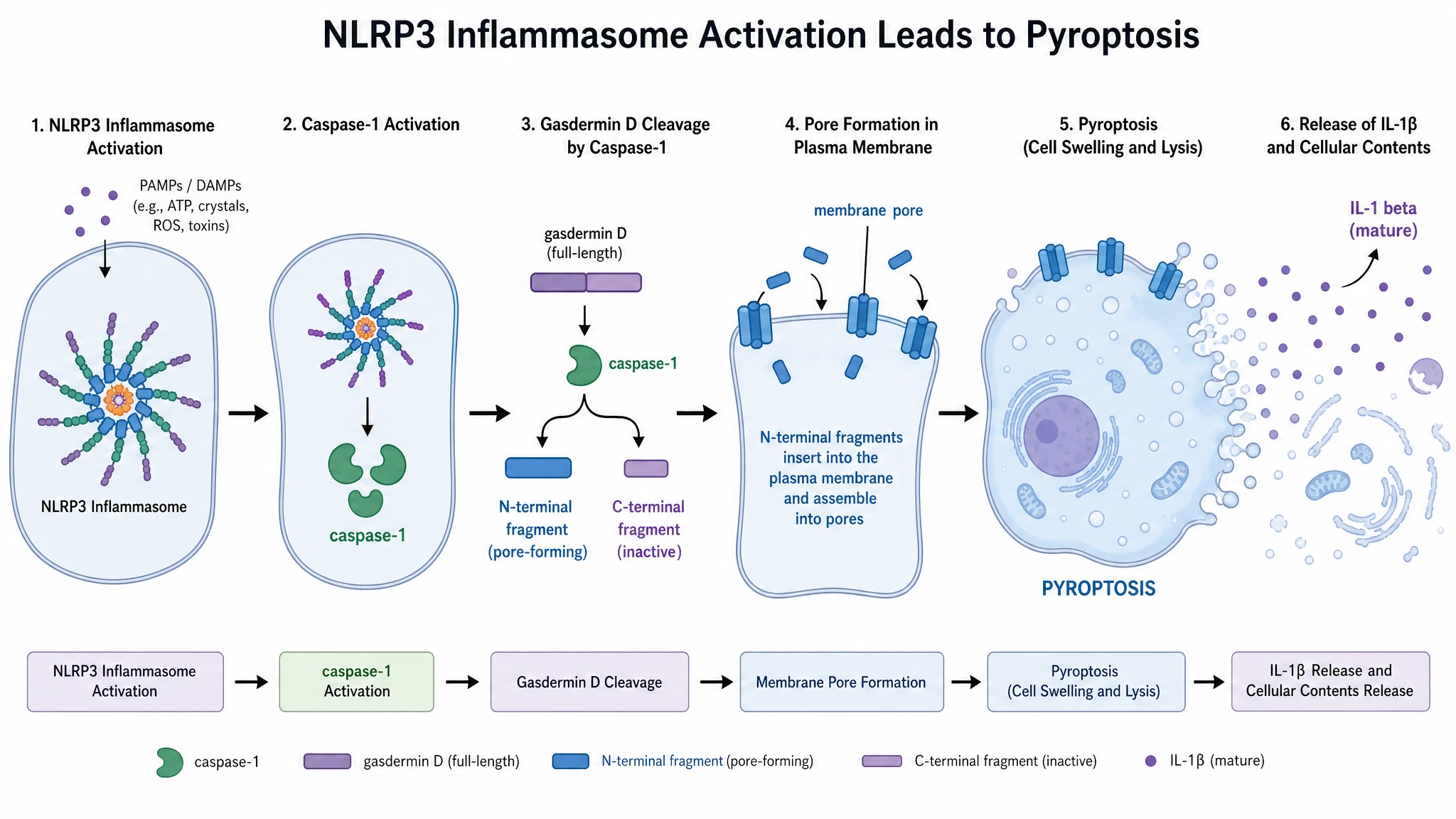
Gasdermin D pores drive release and pyroptosis
Caspase-1 also cleaves gasdermin D, freeing an N-terminal fragment that inserts into the inner leaflet of the plasma membrane and oligomerises into pores roughly 10-20 nm across. Mature IL-1 beta exits through them. Sustained pore formation causes osmotic swelling and, with NINJ1-mediated membrane rupture, lytic pyroptotic death that spills the remaining cytosolic contents, including DAMPs that recruit and prime neighbouring cells.
NLRP3 Inflammasome Pathway— templates & examples
How to make NLRP3 Inflammasome Pathway
Describe your figure
Tell SciFig what to draw in plain language — no design tools required.
Generate with SciFig
Get a clean, publication-ready figure that matches your description in seconds.
Edit & export
Vectorize it into editable SVG, relabel everything, and export for your paper, poster, or slides.
Related searches
- nlrp3 inflammasome pathway
- inflammasome pathway
- nlrp3 inflammasome activation
- nlrp3 inflammasome signaling pathway
- inflammasome diagram
- nlrp3 inflammasome assembly
- il-1 beta inflammasome pathway
Make figures for
Related pages
Immunology Templates
Browse more Immunology templates in SciFig's library.
All Templates
Browse every template page in SciFig's library.
Graphical Abstract Maker
Summarize your study in one panel
Scientific Poster Generator
Design a conference research poster
AI Scientific Image Generator
Generate research figures from text
Conceptual Framework Generator
Map variables and their relationships
Frequently Asked Questions
Common questions about NLRP3 Inflammasome Pathway.

Ready to publish?
Make your own NLRP3 Inflammasome Pathway in minutes.
Start for freeFree to start · No credit card required · Built for researchers