
Membrane Protein Functions
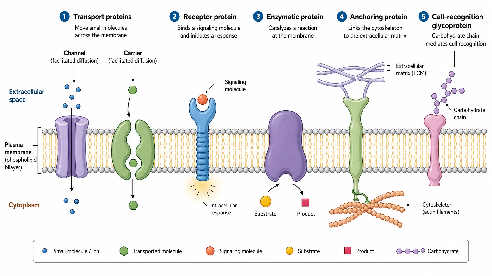
A labeled diagram of the functions of membrane proteins in the plasma membrane — transport, signaling, enzymatic, anchoring, and recognition — ready to relabel and export.
Updated

Start with up to 150 free credits|No credit card required
Get up to 350 free credits on day one when you join through an invite.
What is Membrane Protein Functions?
A membrane protein functions diagram illustrates the main jobs proteins perform in the plasma membrane. It shows transport proteins (channels and carriers) moving molecules across the lipid bilayer, receptor proteins binding signaling molecules, enzymatic proteins catalyzing reactions, anchoring proteins linking the cytoskeleton and matrix, and cell-recognition glycoproteins acting as identity markers. With SciFig you generate a publication-ready membrane figure you can relabel and export.
Why This Diagram Gets Drawn
- The roles are defined by position in the bilayer, and position is spatial information that prose handles badly.
- A single panel lets a reader compare a channel, a carrier, and a pump side by side, where the mechanistic differences are otherwise blurred.
- It anchors the fluid-mosaic model concretely: the lipid bilayer is the solvent, and the proteins do essentially all of the work.
- Papers on a specific transporter or receptor need one orienting figure showing where that protein sits among the other roles.
- Distinguishing integral from peripheral association is difficult to convey without showing the hydrophobic transmembrane segment.
- Cell-biology teaching returns to this figure constantly; a clean, correctly scaled version replaces several textbook diagrams.
Elements to Include
- Channel proteins — a hydrophilic pore with a selectivity filter, gated by voltage, ligand, or mechanical stress.
- Carrier proteins — alternating-access conformational cycling; facilitated diffusion (GLUT1) and secondary active symport (SGLT1).
- Pumps — primary active transport driven by ATP hydrolysis, with stoichiometry shown (Na⁺/K⁺-ATPase, Ca²⁺-ATPase, H⁺-ATPase).
- Receptors — GPCRs and receptor tyrosine kinases, with the intracellular effector shown so transduction is visible rather than implied.
- Enzymatic proteins — a catalytic site presented to the cytosol or extracellular space; adenylyl cyclase, γ-secretase, alkaline phosphatase.
- Anchoring and junctional proteins — integrin–talin–actin coupling to the cytoskeleton; tight junctions, gap junctions, and desmosomes.
- Recognition glycoproteins — branched extracellular glycans acting as cell-identity markers, plus a clear integral-versus-peripheral contrast.
Where This Figure Is Used
- Introduction figures in structural-biology papers, placing a newly solved transporter or receptor in its functional context.
- Pharmacology reviews, since most drug targets are receptors, channels, or transporters at the cell surface.
- Neuroscience and physiology teaching on membrane potential, where channels, carriers, and pumps must be distinguished.
- Immunology figures relying on surface glycoproteins and MHC as recognition markers.
- Cell-adhesion and mechanotransduction papers showing integrin coupling between matrix and cytoskeleton.
- Lecture slides, exam materials, and textbook chapters covering the functions of membrane proteins as a set.
What the Diagram Has to Get Right
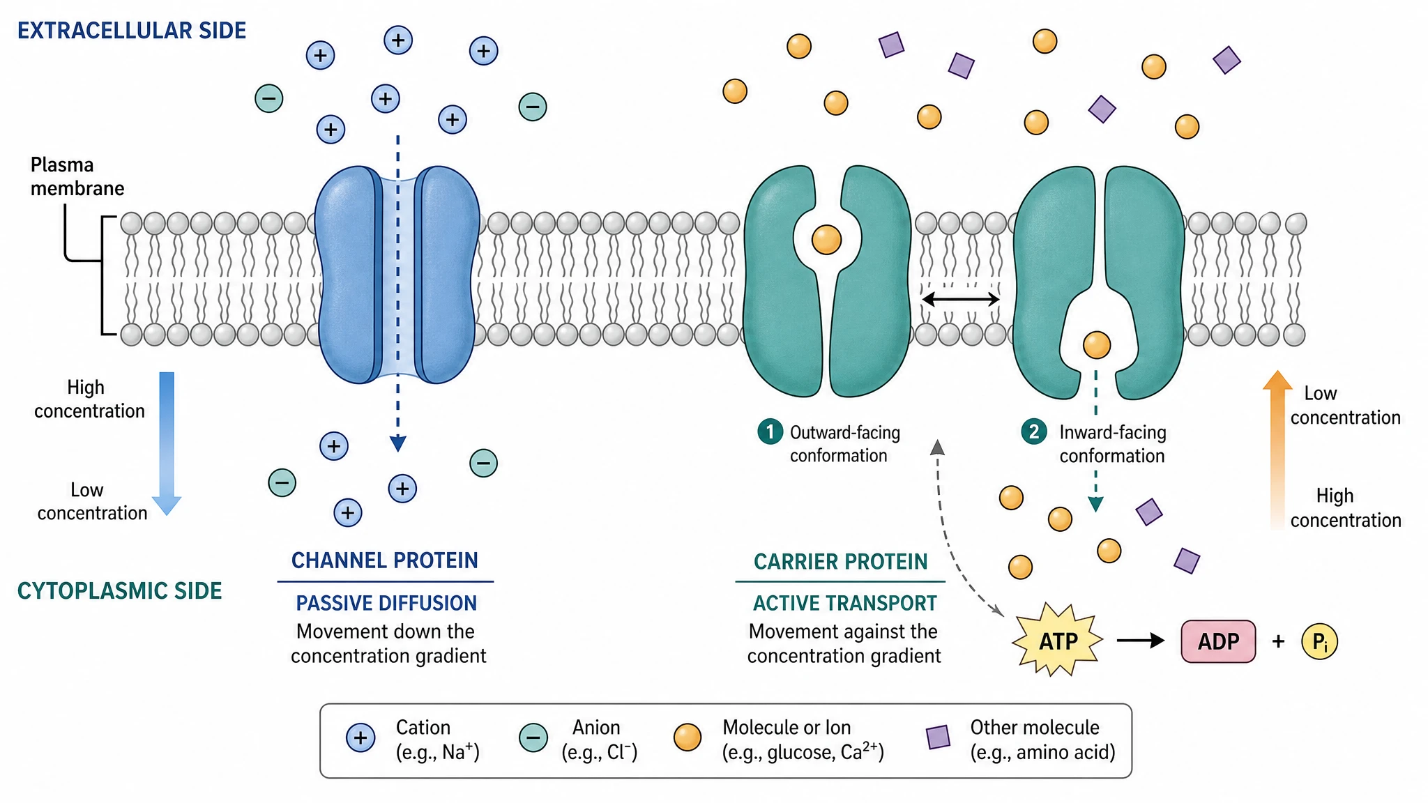
Transport: Channels, Carriers, and Pumps Are Not Interchangeable
Three mechanisms deserve three distinct depictions. Channels form a continuous aqueous pore and move solute down its gradient at near-diffusion rates — aquaporin for water, the selectivity filter of a voltage-gated K⁺ channel for ions. Carriers such as GLUT1 alternate between outward- and inward-open conformations, one substrate at a time. Pumps hydrolyse ATP to work against the gradient: Na⁺/K⁺-ATPase exports three Na⁺ and imports two K⁺ per ATP.
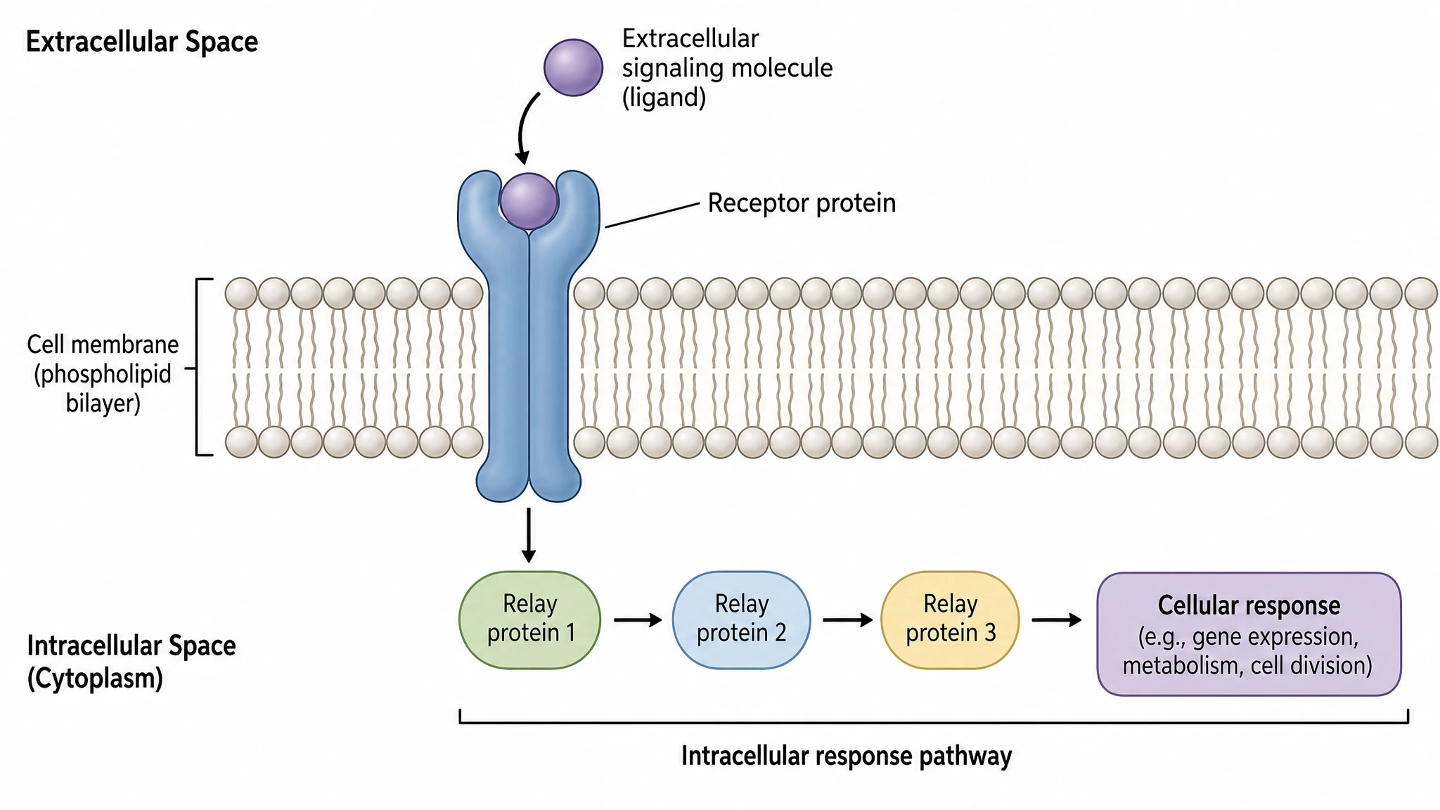
Receptors Transduce Signals Across the Bilayer
A signalling receptor converts extracellular ligand binding into an intracellular event without the ligand crossing the lipid bilayer. Draw the two dominant classes: GPCRs, whose seven-helix bundle catalyses GDP-to-GTP exchange on the Gα subunit and activates adenylyl cyclase or phospholipase C; and receptor tyrosine kinases such as EGFR, which dimerise, trans-autophosphorylate cytoplasmic tyrosines, and recruit SH2-domain adaptors into the MAPK cascade.
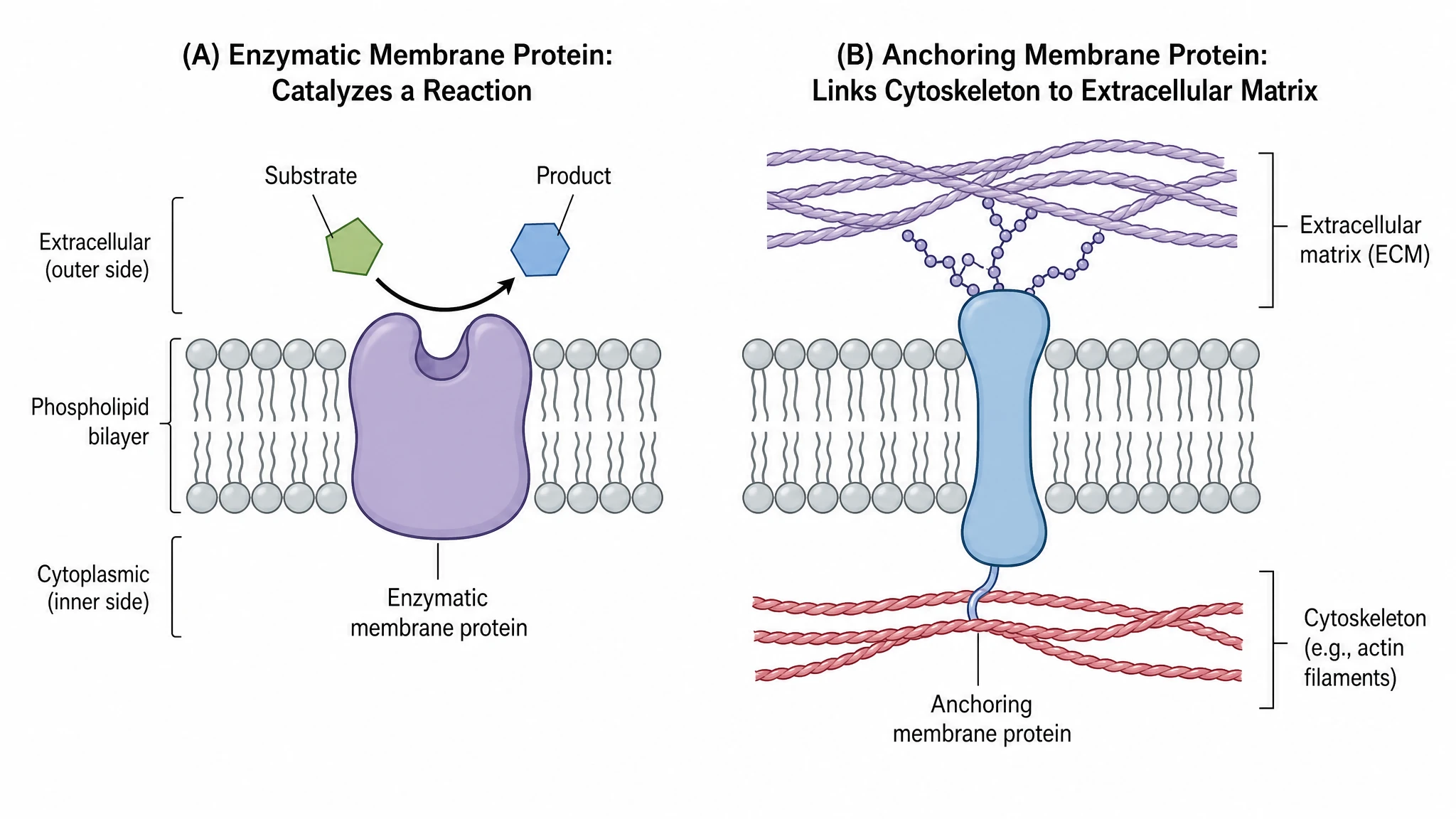
Catalysis, Anchoring, and Cell Junctions
Some bilayer-embedded proteins are enzymes with the active site at the membrane surface — adenylyl cyclase, γ-secretase, alkaline phosphatase. Others are structural: integrins bind extracellular fibronectin and connect through talin and vinculin to the actin cytoskeleton, transmitting mechanical force. Junctional proteins are a third role: claudins and occludins seal tight junctions, connexin hexamers form gap-junction channels, and cadherins hold desmosomes together.
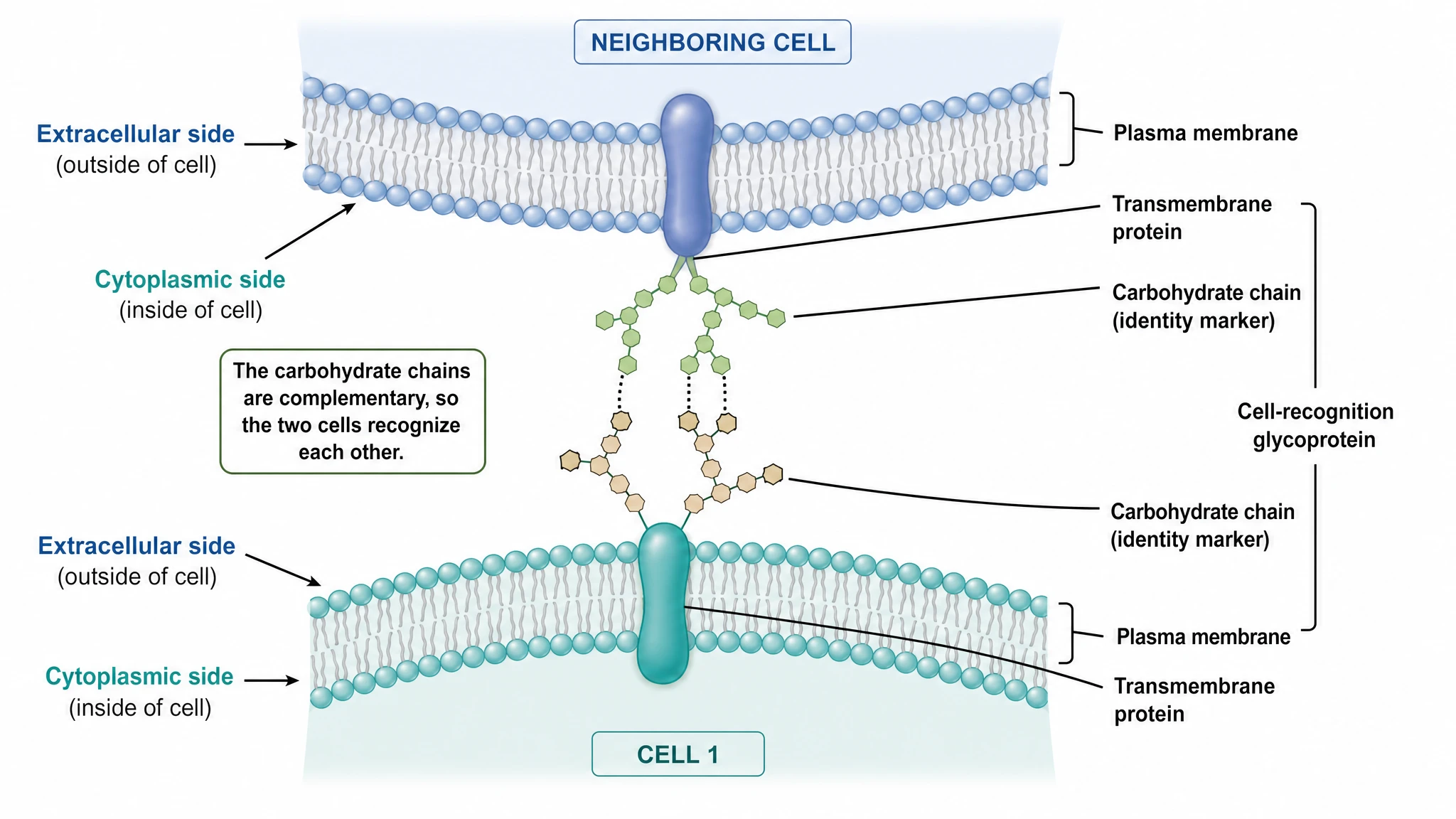
Recognition Glycoproteins and Bilayer Topology
Cell-surface identity is carried by N- and O-linked glycans on extracellular loops: ABO blood-group determinants, MHC class I and II presenting peptide to T cells, and selectin–carbohydrate binding that slows rolling leukocytes. A rigorous figure also distinguishes integral proteins — single-pass or multi-pass hydrophobic α-helices of roughly twenty residues, or β-barrels — from peripheral proteins bound electrostatically or through a GPI anchor.
Membrane Protein Functions— templates & examples
How to make Membrane Protein Functions
Describe your figure
Tell SciFig what to draw in plain language — no design tools required.
Generate with SciFig
Get a clean, publication-ready figure that matches your description in seconds.
Edit & export
Vectorize it into editable SVG, relabel everything, and export for your paper, poster, or slides.
Related searches
- functions of membrane proteins
- membrane protein functions diagram
- types of membrane proteins
- transport proteins membrane
- receptor proteins cell membrane
- integral membrane proteins
- cell recognition proteins
- enzymatic membrane proteins
- anchoring proteins cytoskeleton
- channel and carrier proteins
Make figures for
Related pages
Cell Biology Templates
Browse more Cell Biology templates in SciFig's library.
All Templates
Browse every template page in SciFig's library.
Graphical Abstract Maker
Summarize your study in one panel
Scientific Poster Generator
Design a conference research poster
AI Scientific Image Generator
Generate research figures from text
Conceptual Framework Generator
Map variables and their relationships
Frequently Asked Questions
Common questions about Membrane Protein Functions.

Ready to publish?
Make your own Membrane Protein Functions in minutes.
Start for freeFree to start · No credit card required · Built for researchers