
Leaf Cross-Section (3D)
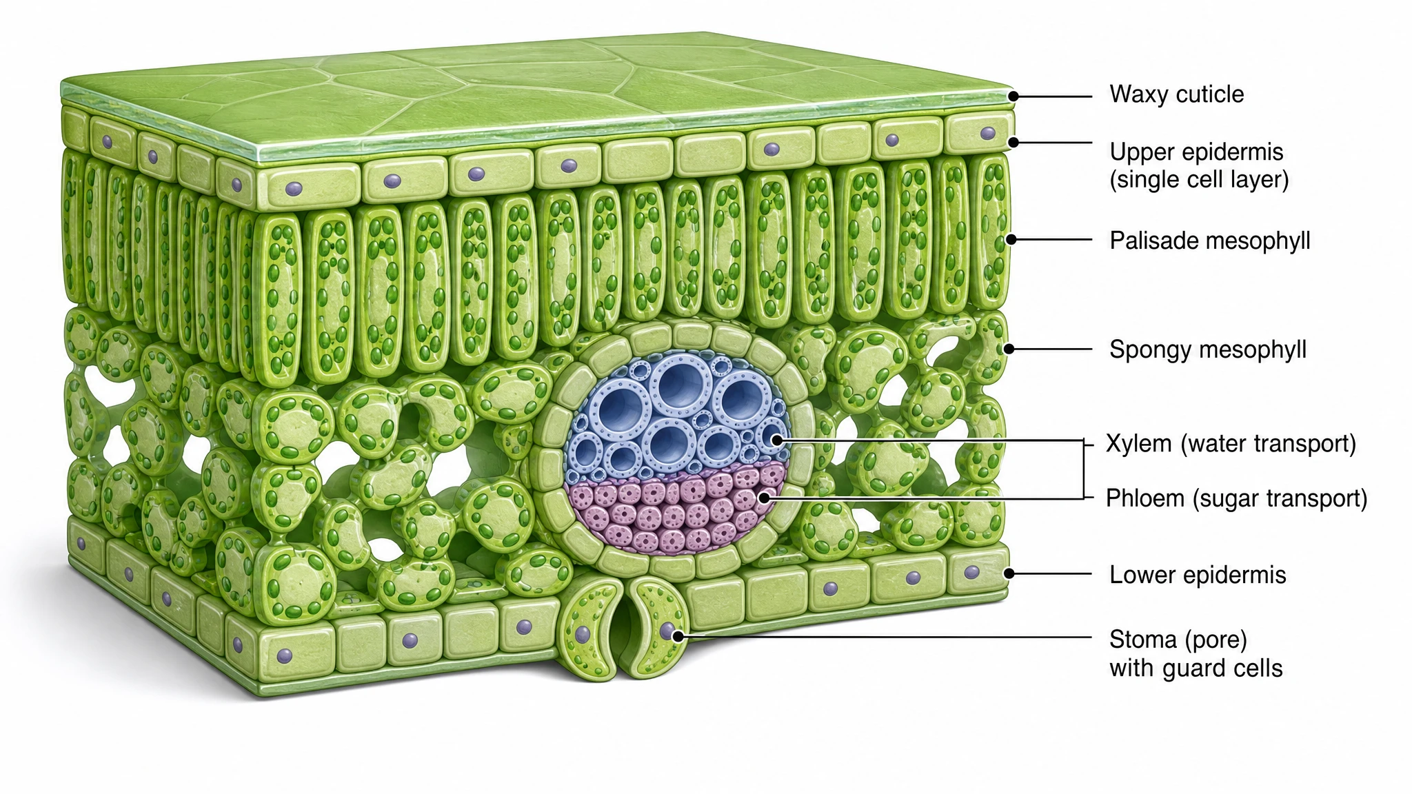
A 3D, labeled cross section of a leaf showing the internal tissue layers — from waxy cuticle and epidermis through the mesophyll to the vascular bundle and stomata.
Updated

Start with up to 150 free credits|No credit card required
Get up to 350 free credits on day one when you join through an invite.
What is Leaf Cross-Section (3D)?
A leaf cross section is a cut-through view of a leaf that reveals its internal tissue layers from top to bottom. A labeled 3D diagram shows the waxy cuticle and upper epidermis, the palisade mesophyll where most photosynthesis occurs, the spongy mesophyll with air spaces for gas exchange, the vascular bundle of xylem and phloem, and the lower epidermis with stomata and guard cells. With SciFig you describe the leaf anatomy and generate a clean, labeled cross section you can relabel and export.
Why draw the internal anatomy instead of the outline
- Function follows position. Palisade cells are packed directly beneath the illuminated upper surface; stomata are concentrated on the shaded lower surface where evaporative demand is lower. Only a cut-through view makes that arrangement arguable.
- Photosynthesis and gas exchange are usually taught as separate topics. The transverse view shows them sharing a single continuous internal air space.
- Comparative anatomy needs a baseline. Kranz anatomy in C4 grasses, sunken stomata and multi-layered epidermis in xerophytes, and thin palisade in shade leaves are all deviations from this standard plan, and only read as deviations against it.
- Micrographs of real material are noisy and stain-dependent. A schematic assigns tissue identities that a stained slide only implies.
- Pathogen entry, herbicide uptake and pollutant deposition are all routed through the cuticle and the stomatal pore, so the barrier layers have to be explicit for those arguments to work.
- Water delivery ends and sugar export begins in the same vascular bundle — a fact no external drawing of a leaf can convey.
Tissue layers to label
- Cuticle — a hydrophobic cutin and wax layer secreted by the epidermis; transparent to light, and the principal barrier to uncontrolled water loss. Thicker in sun leaves and in arid-climate species.
- Upper (adaxial) epidermis — a single layer of tightly packed cells with few or no chloroplasts; protective, and transparent enough to pass light to the tissue below.
- Palisade mesophyll — elongated cells stacked perpendicular to the surface with the highest chloroplast density in the leaf; the main site of carbon fixation.
- Spongy mesophyll — irregularly shaped cells separated by large intercellular air spaces, giving the high surface-to-volume ratio that lets CO2 diffuse to every chloroplast.
- Vascular bundle — xylem on the adaxial side carrying water and minerals up from the root, phloem on the abaxial side exporting sucrose, both enclosed by a bundle sheath.
- Lower (abaxial) epidermis — the surface bearing most of the stomata in a typical dorsiventral leaf.
- Stomata and guard cells — the pore, the paired guard cells that control it, and the substomatal cavity that connects it to the internal air space.
Where this diagram is used
- Introductory botany and plant physiology teaching, where the tissue layers anchor the entire photosynthesis unit.
- C3, C4 and CAM comparisons, where Kranz anatomy and bundle-sheath chloroplasts are drawn against the standard dorsiventral plan.
- Drought and xerophyte adaptation studies, illustrating thickened cuticle, sunken stomata and reduced stomatal density.
- Stomatal density and conductance work, including palaeoclimate reconstructions from fossil leaf material.
- Plant pathology, tracing fungal and bacterial entry through the stomatal pore or through breaches in the cuticle.
- Microscopy papers, where a schematic sits beside a stained transverse section to name what the reader is looking at.
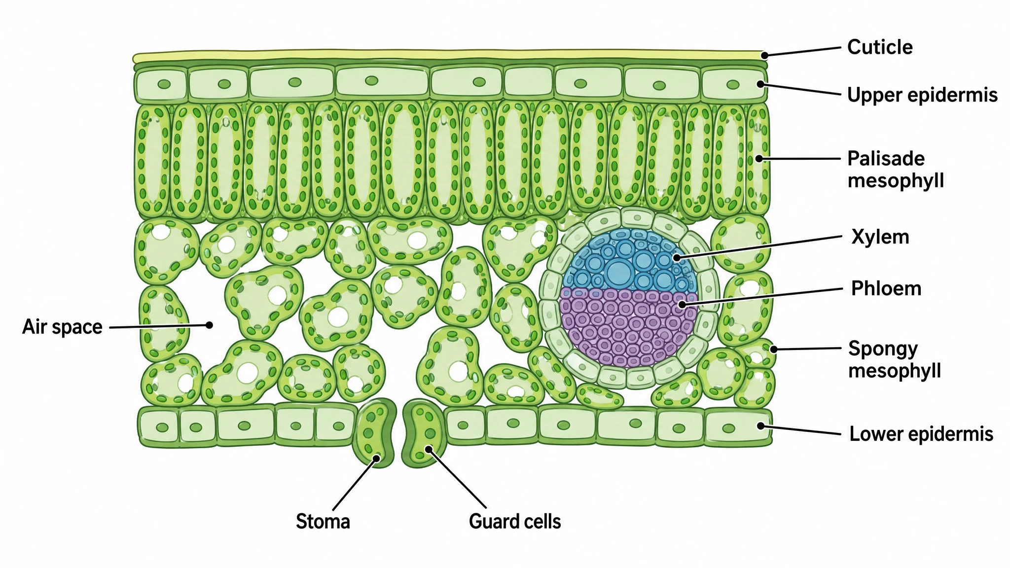
What a labeled transverse view of leaf tissue actually shows, layer by layer.
Tissue layers from cuticle down to lower epidermis
Read the section top to bottom: a waxy cuticle of cutin secreted by the epidermis; the upper epidermis itself, a single transparent layer with few or no chloroplasts; the palisade mesophyll, one to three ranks of columnar, chloroplast-dense cells; the spongy mesophyll with its air spaces; and the lower epidermis carrying the stomata. Labeling every layer in one figure is what makes the anatomy teachable.
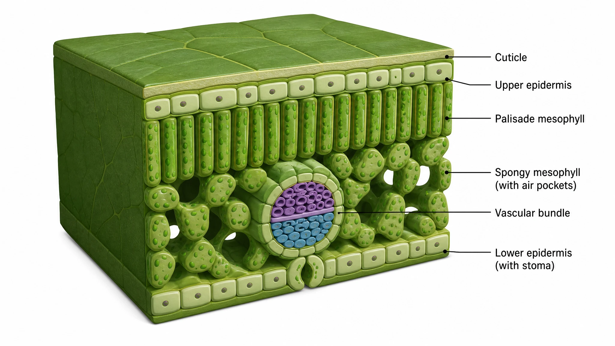
A 3D cutaway makes the air spaces legible
A flat drawing implies that the intercellular gaps of the spongy layer are isolated pockets. They are not: they form a connected volume that runs laterally through the mesophyll and opens to the atmosphere at the substomatal cavity. A cutaway with depth shows that continuity, and it also shows that palisade cells are columns rather than the rectangles a two-dimensional view suggests.
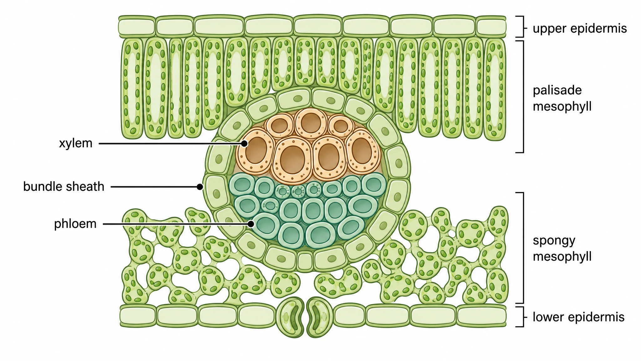
Xylem above, phloem below, bundle sheath around
In the midrib and the minor veins the bundle keeps a fixed orientation. Xylem sits adaxially, toward the upper surface, delivering water and dissolved minerals from the root through vessels and tracheids. Phloem sits abaxially, exporting sucrose from the photosynthetic tissue through sieve tube elements. A bundle sheath of tightly packed parenchyma encircles both. The orientation is reversed in stems, and it is the detail students most often get wrong.
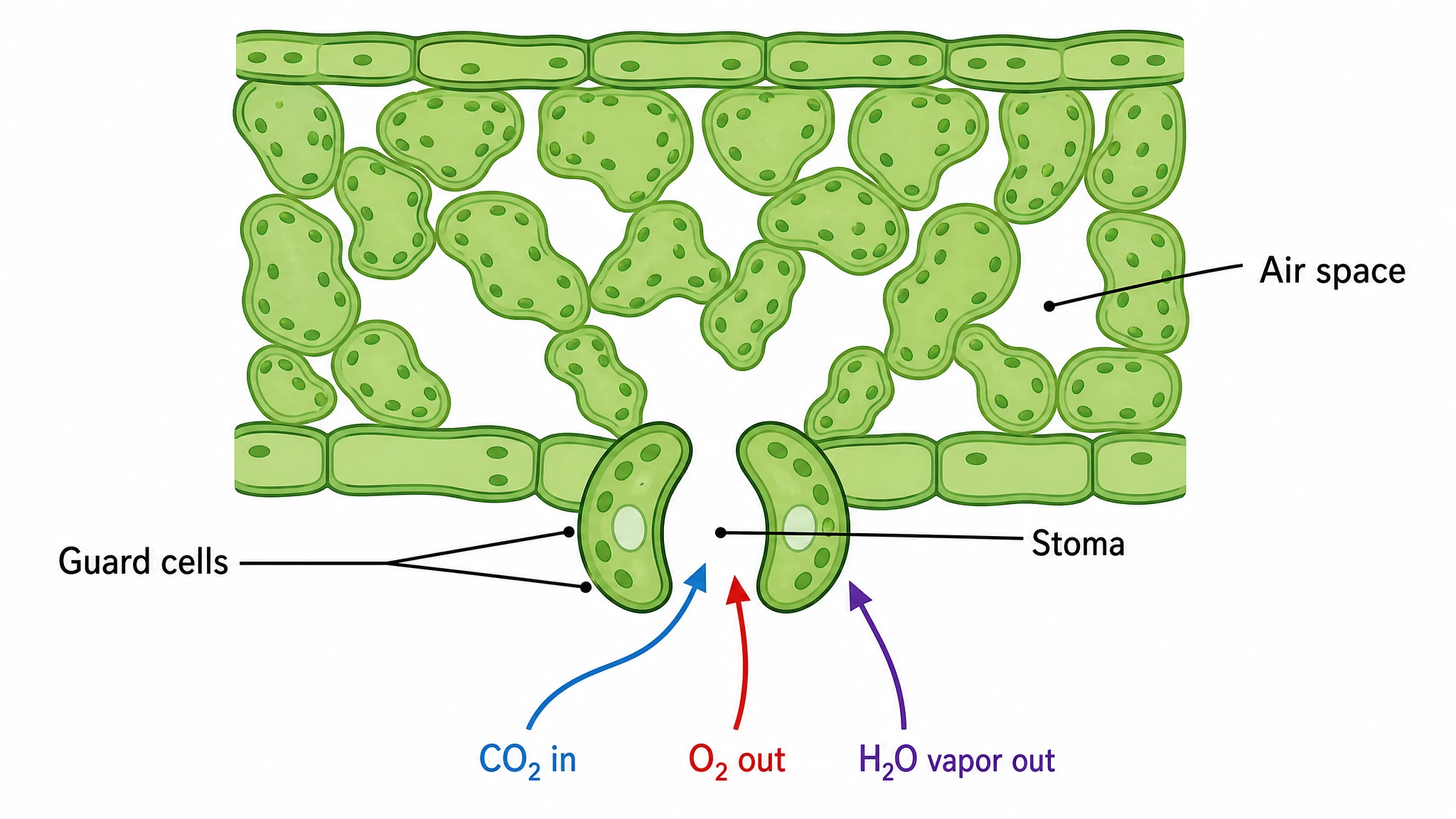
Guard cells set the gas-exchange trade-off
Each stoma is a pore flanked by two kidney-shaped guard cells whose walls are thickened on the inner face. Potassium influx draws water in osmotically, the cells swell, and the unequal wall thickening bows them apart to open the pore. Carbon dioxide enters and water vapour leaves through the same aperture, so the plant cannot take up carbon without paying in water — the trade-off the figure should make visible.
Leaf Cross-Section (3D)— templates & examples
How to make Leaf Cross-Section (3D)
Describe your figure
Tell SciFig what to draw in plain language — no design tools required.
Generate with SciFig
Get a clean, publication-ready figure that matches your description in seconds.
Edit & export
Vectorize it into editable SVG, relabel everything, and export for your paper, poster, or slides.
Related searches
- cross section of a leaf
- cross section a leaf labeled diagram
- leaf cross section
- 3d cross section diagram
- simple cross section of leaf
- cross section of a leaf diagram
- 3d diagram of leaf cross section
- cross section of a leaf structure
- what is a leaf cross section
- cross section of leaf model with cellular system
Make figures for
Related pages
Botany Templates
Browse more Botany templates in SciFig's library.
All Templates
Browse every template page in SciFig's library.
Graphical Abstract Maker
Summarize your study in one panel
Scientific Poster Generator
Design a conference research poster
AI Scientific Image Generator
Generate research figures from text
Conceptual Framework Generator
Map variables and their relationships
Frequently Asked Questions
Common questions about Leaf Cross-Section (3D).

Ready to publish?
Make your own Leaf Cross-Section (3D) in minutes.
Start for freeFree to start · No credit card required · Built for researchers