
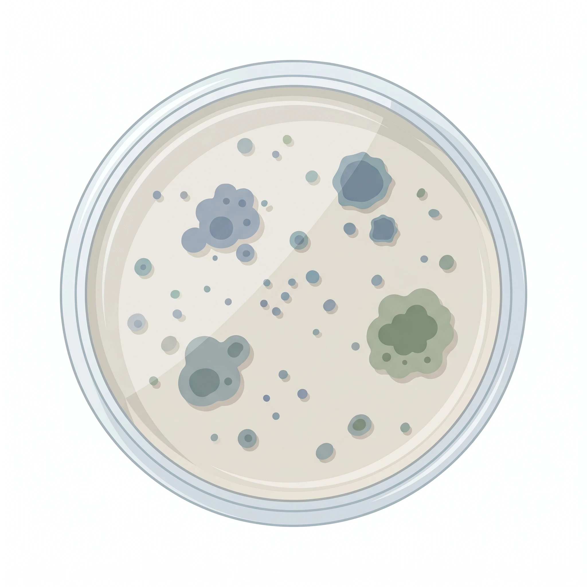
Petri Dish With Cells
A clean, editable petri dish with cells for microbiology figures, posters, and slides — ready to relabel and export in any format.
Updated

Start with up to 150 free credits|No credit card required
Get up to 350 free credits on day one when you join through an invite.
What is Petri Dish With Cells?
A petri dish with cells is a common microbiology icon used to show cultured cells, bacterial colonies, or growth media in a shallow, lidded dish. It appears in lab protocols, graphical abstracts, and teaching figures whenever a culture step needs a clear visual. With SciFig you describe the dish you need in plain language and generate a clean, editable petri dish — top view or angled, empty or seeded — ready to relabel and export.
Why figures use a drawn culture plate instead of a photo
- A photograph of a plate carries lens glare, condensation, and background clutter that survives neither greyscale conversion nor a 40 mm single-column figure; a vector drawing stays legible at any reduction.
- The dish is a standardised visual token: readers parse a shallow circle with a rim as "culture step" instantly, which lets a workflow schematic skip explanatory text.
- Drawing the plate lets you show a state that no camera captured — a seeding density, a colony count, or a treatment condition that exists only as an experimental design.
- Consistent geometry across panels matters: if panel A shows a 10 cm dish and panel B shows a 35 mm dish, the drawing must encode that difference rather than leave it to the caption.
- Journals increasingly require that schematic elements be distinguishable from data; a stylised plate signals "illustration", not "result", and avoids the image-integrity flags a decorative micrograph attracts.
What to draw and label on the plate
- Lid and base as separate strokes: the lid is slightly larger in diameter and overhangs the base, and a small offset between them is what makes a top view read as a lidded vessel rather than a flat disc.
- The agar or medium layer — a filled band inset from the wall, drawn with a visible meniscus in side view. Its depth (typically ~4 mm for a 10 cm plate) distinguishes an agar plate from a liquid culture dish.
- The biological content itself, and it must match the claim: discrete circular colonies for a bacterial streak or spread plate; a confluent or subconfluent monolayer stipple for adherent mammalian cells; a lawn for a phage plaque assay.
- Choose one viewpoint and stay with it. A true top view (concentric circles) is best for colony counting and CFU panels; a 20–30° angled or side view is better when you need to show medium depth, a lid being lifted, or liquid handling.
- Optional annotations that carry information: quadrant streak lines, a grid for counting, a plate label or barcode strip, and a scale cue such as the plate diameter (35 / 60 / 100 mm).
- Keep the colony marks irregular in placement — evenly spaced dots on a grid read as decoration and mislead readers about plating density.
Where the drawn dish appears
- Method schematics for isolation and culture: sample → enrichment broth → streak → single-colony pick, with each plate showing the appropriate colony morphology.
- Antimicrobial and susceptibility figures: disc diffusion plates with zones of inhibition, MIC agar dilution series, or checkerboard assay layouts.
- Cell-culture workflow panels in graphical abstracts — passage, seeding, transfection, and treatment timelines where the dish anchors each time point.
- Teaching slides and SOP documents for aseptic technique, where the lid position (on, ajar, off) is itself the instructional content.
- Screening and phenotype figures: colony PCR pipelines, blue/white selection, yeast spot dilution assays, and CFU quantification panels.
Petri Dish With Cells— templates & examples
How to make Petri Dish With Cells
Describe your figure
Tell SciFig what to draw in plain language — no design tools required.
Generate with SciFig
Get a clean, publication-ready figure that matches your description in seconds.
Edit & export
Vectorize it into editable SVG, relabel everything, and export for your paper, poster, or slides.
Related searches
- petri dish with cells
- petri dish diagram
- petri dish png
- petri dish illustration
- petri plate clipart
- cells in a dish
- petri dish graphic
- culture plate icon
- agar plate diagram
- microbiology culture plate
Make figures for
Related pages
Microbiology Icons
Browse more Microbiology icons in SciFig's library.
All Icons
Browse every icon page in SciFig's library.
AI Scientific Image Generator
Generate research figures from text
Graphical Abstract Maker
Summarize your study in one panel
Text to Diagram Generator
Turn text into a diagram
Scientific Poster Generator
Design a conference research poster
Frequently Asked Questions
Common questions about Petri Dish With Cells.

Ready to publish?
Make your own Petri Dish With Cells in minutes.
Start for freeFree to start · No credit card required · Built for researchers