
Aortic Valve Bicuspid
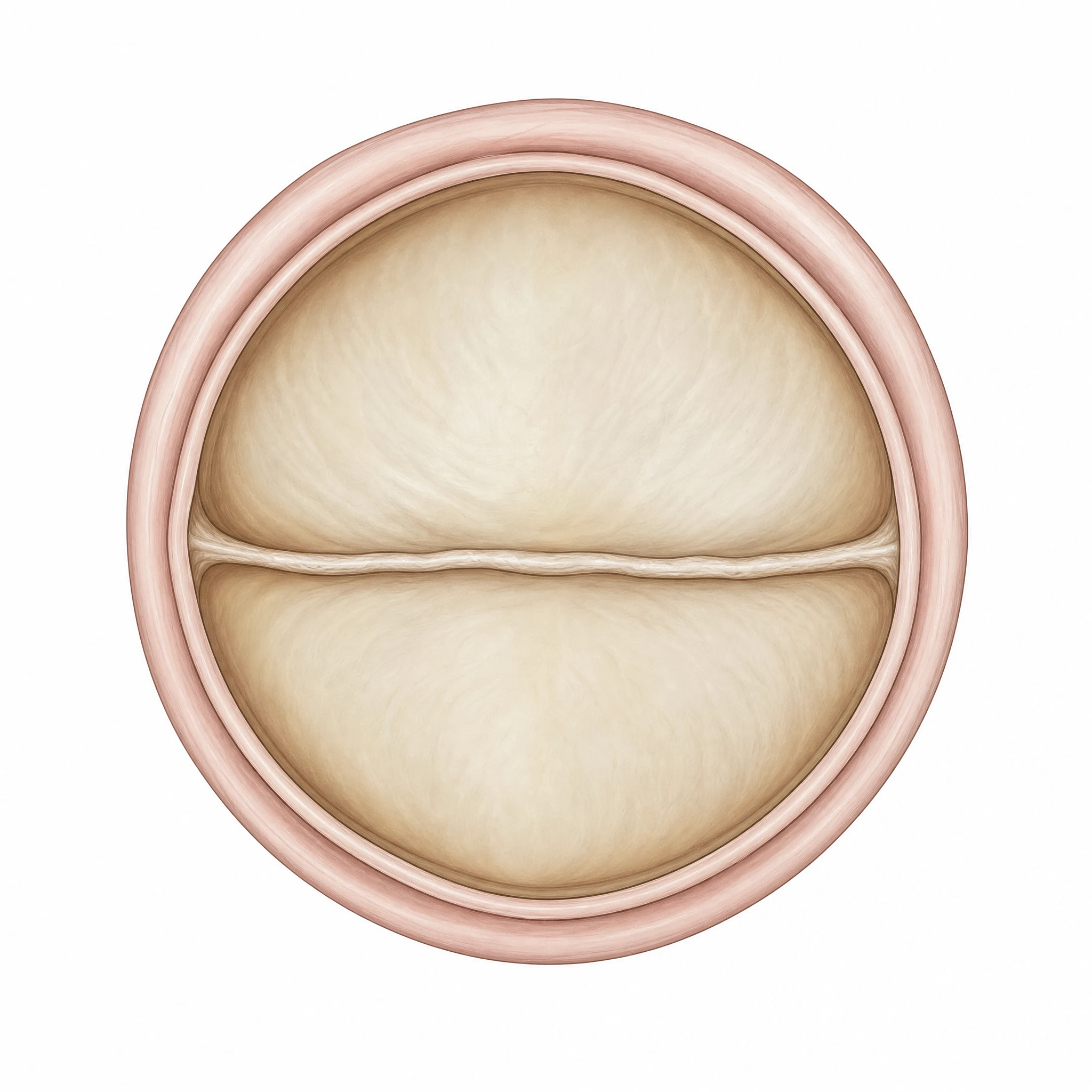
A clean, editable bicuspid aortic valve showing two leaflets instead of the normal three — ready for cardiology figures, posters, and teaching slides.
Updated

Start with up to 150 free credits|No credit card required
Get up to 350 free credits on day one when you join through an invite.
What is Aortic Valve Bicuspid?
A bicuspid aortic valve is a congenital heart valve that has two leaflets (cusps) instead of the normal three, so it opens and closes through a single fused commissure. It is a common cardiology icon used in figures on aortic stenosis, valve disorders, and congenital heart disease, often shown alongside a normal tricuspid valve for comparison. With SciFig you describe the valve you need in plain language and generate a clean, editable bicuspid aortic valve, ready to relabel and export.
Why the malformed valve is drawn beside the normal one
- The abnormality is only visible as a contrast. A two-leaflet valve drawn alone is just a shape; drawn next to the three-cusp arrangement, the fused commissure and the elliptical rather than triangular orifice become self-evident.
- It is the most common congenital cardiac malformation, affecting roughly 1–2% of the population, and it accounts for a large share of aortic stenosis requiring intervention — so it appears constantly in cardiology figures and needs a reliable visual convention.
- Morphology drives prognosis: which commissure is fused, and whether a raphe is present, predicts the pattern of stenosis versus regurgitation and the risk of aortopathy. A drawing that omits the raphe omits the clinically decisive feature.
- Echocardiographic stills are grainy and orientation-dependent; a clean short-axis illustration is what makes the leaflet arrangement teachable.
- Consistency with the accepted classification matters, because a figure that invents its own leaflet naming cannot be compared with the surgical literature.
Anatomy to render and label
- The normal tricuspid reference: right coronary, left coronary, and non-coronary cusps, with the right and left coronary ostia arising from their respective sinuses of Valsalva — drawn in short axis, the closed valve makes the classic three-radial-line (Mercedes) pattern.
- The two-leaflet configuration itself: one conjoined leaflet formed by fusion of two cusps plus one non-fused leaflet, giving a fish-mouth systolic orifice rather than a triangular one.
- The raphe — the fibrous ridge marking the fused commissure on the conjoined leaflet. Its presence (and its extent to the free margin) distinguishes a true two-cusp valve from a fused-raphe variant and belongs in the label set.
- The Sievers classification, which the drawing should be able to express: Type 0 (no raphe, two symmetric cusps), Type 1 (one raphe — subtypes L-R, R-N, N-L by which commissure is fused, L-R being by far the most common), Type 2 (two raphes).
- Both systolic (open) and diastolic (closed) states. The reduced, eccentric opening area in systole is what produces the stenotic gradient; incomplete coaptation in diastole is what produces regurgitation.
- The associated aorta, when the figure concerns aortopathy: annulus, sinuses, sinotubular junction, and ascending aorta, since dilatation of the ascending segment or root is a companion finding requiring surveillance.
Where the illustration appears
- Cardiology and cardiac-surgery figures on valvular stenosis and regurgitation — natural history, gradient progression, and intervention thresholds.
- Echocardiography teaching material: parasternal short-axis views, planimetry of the orifice, and the acquisition planes that reveal a raphe.
- Intervention and surgical schematics — surgical AVR, the Ross procedure, valve repair, and the specific anatomical considerations that make TAVI in this morphology non-standard (elliptical annulus, asymmetric calcification, raphe calcification).
- Aortopathy and genetics figures: association with ascending aortic aneurysm, coarctation, and Turner syndrome; NOTCH1 and familial clustering.
- Registry and imaging-cohort papers where the Sievers subtype is the stratifying variable and each arm needs its own diagram.
- Patient-education and consent material, where the two-versus-three leaflet contrast is the entire explanation.
Aortic Valve Bicuspid— templates & examples
How to make Aortic Valve Bicuspid
Describe your figure
Tell SciFig what to draw in plain language — no design tools required.
Generate with SciFig
Get a clean, publication-ready figure that matches your description in seconds.
Edit & export
Vectorize it into editable SVG, relabel everything, and export for your paper, poster, or slides.
Related searches
- bicuspid aortic valve
- bicuspid aortic valve diagram
- two leaflet aortic valve
- bicuspid aortic valve png
- bicuspid vs tricuspid aortic valve
- aortic valve icon
- congenital heart valve diagram
- aortic valve leaflets diagram
- heart valve disorder figure
- valve disorder gif
Make figures for
Related pages
Cardiology Icons
Browse more Cardiology icons in SciFig's library.
All Icons
Browse every icon page in SciFig's library.
AI Scientific Image Generator
Generate research figures from text
Graphical Abstract Maker
Summarize your study in one panel
Text to Diagram Generator
Turn text into a diagram
Scientific Poster Generator
Design a conference research poster
Frequently Asked Questions
Common questions about Aortic Valve Bicuspid.

Ready to publish?
Make your own Aortic Valve Bicuspid in minutes.
Start for freeFree to start · No credit card required · Built for researchers