CAR-T Mechanism Illustration for EHA 2026 Posters
Draw publication-ready CAR-T mechanism diagrams for EHA 2026 posters: 5 visual components, 4 CAR generations, BiTE family, and copy-paste AI prompts.
You are five days from the EHA poster print deadline, and your CAR-T mechanism figure still looks like a high-school biology textbook. The scFv has three domains where it should have two. The CD3ζ chain is pointing the wrong way. Your second-generation construct and your third-generation construct look indistinguishable. You have burned 90 minutes in Adobe Illustrator nudging arrows, and the figure still does not convey the immunological synapse you actually study.
This is the moment that breaks most poster preparation timelines for hematology researchers presenting cellular immunotherapy work. CAR-T is the single most searched visual category in EHA's Tier 1 Gene therapy and cellular immunotherapy topic — and the most difficult to render correctly. This guide walks through the five visual components every CAR construct figure needs, the four generations of CAR design, the bispecific antibody family that dominates current trials, and the AI-assisted workflow that compresses 8 hours of Illustrator labor into 30 minutes of structured prompting.
Transparency note: Illustrations in this article were generated with SciFig AI and reviewed by the author for scientific accuracy. Cited claims link to peer-reviewed sources, FDA labels, and ASH/EHA educational materials.
1. Why CAR-T Mechanism Diagrams Are EHA's Most Searched Visual
The problem is that "CAR-T mechanism" is not one figure — it is at least four overlapping figures: the CAR construct itself, the immunological synapse, the manufacturing workflow, and the adverse-event pathophysiology. Reviewers expect all four. Most poster authors only draft one or two well and let the others fall flat.
This guide fixes that by giving you the visual vocabulary for each, plus the SciFig prompts you can copy directly.
2. Anatomy of a CAR-T Mechanism Figure: 5 Visual Components Every Construct Needs
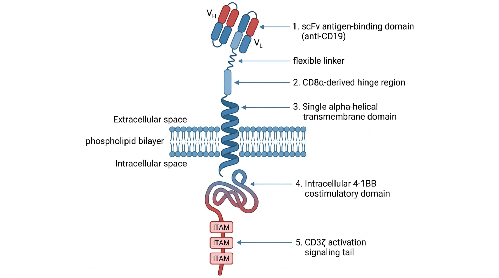
Every CAR construct figure has the same five visual components from outside the cell to inside. Get any one wrong and an experienced reviewer will spot the error within seconds.
- scFv antigen-binding domain — Two variable domains (V_H and V_L) joined by a flexible linker, drawn outside the T-cell membrane. Common error: drawing one domain instead of two, or three instead of two.
- Hinge region — A flexible stalk (typically derived from CD8α or IgG4) connecting scFv to the transmembrane domain. Often omitted or drawn too short.
- Transmembrane (TM) domain — A single alpha-helical pass through the phospholipid bilayer. Must clearly traverse both membrane leaflets, not float above or only pierce one.
- Costimulatory domain — Intracellular, either 4-1BB (CD137) or CD28. This is what distinguishes second- and third-generation constructs from each other.
- CD3ζ activation domain — Intracellular, contains three ITAM (Immunoreceptor Tyrosine-based Activation Motif) signaling motifs that fire upon antigen binding. Common error: drawing two ITAMs instead of three, or showing the CD3ζ chain pointing the wrong way (extracellular).
When you copy a SciFig prompt below, the model produces a starter image that gets these five components topologically correct most of the time. Your remaining work — using SciFig's vector canvas — is renaming domains to match your specific construct (anti-BCMA vs anti-CD19 vs anti-GPRC5D), swapping costimulatory domains, and adjusting label fonts to match your poster typography.
3. The 4 Generations of CAR Constructs: Visualizing 4-1BB vs CD28 Costimulation
Showing the evolution of CAR design across four generations on a single panel is one of the most reviewer-impressive figures you can include — and it is also where AI generators most often confuse 4-1BB and CD28 positions or scramble the order of generations.
- Generation 1 — scFv + CD3ζ only. No costimulation. Largely abandoned clinically due to poor persistence in patients.
- Generation 2 — Adds a single costimulatory domain: either CD28 (Yescarta, axicabtagene ciloleucel) or 4-1BB (Kymriah, tisagenlecleucel). The dominant clinical scaffold today.
- Generation 3 — Dual costimulation: both 4-1BB and CD28 in series. Trials underway but no approvals as of 2026.
- Generation 4 ("armored CAR") — Adds an inducible cytokine secretion module (typically IL-12 or IL-18). Designed to remodel the tumor microenvironment from inside.
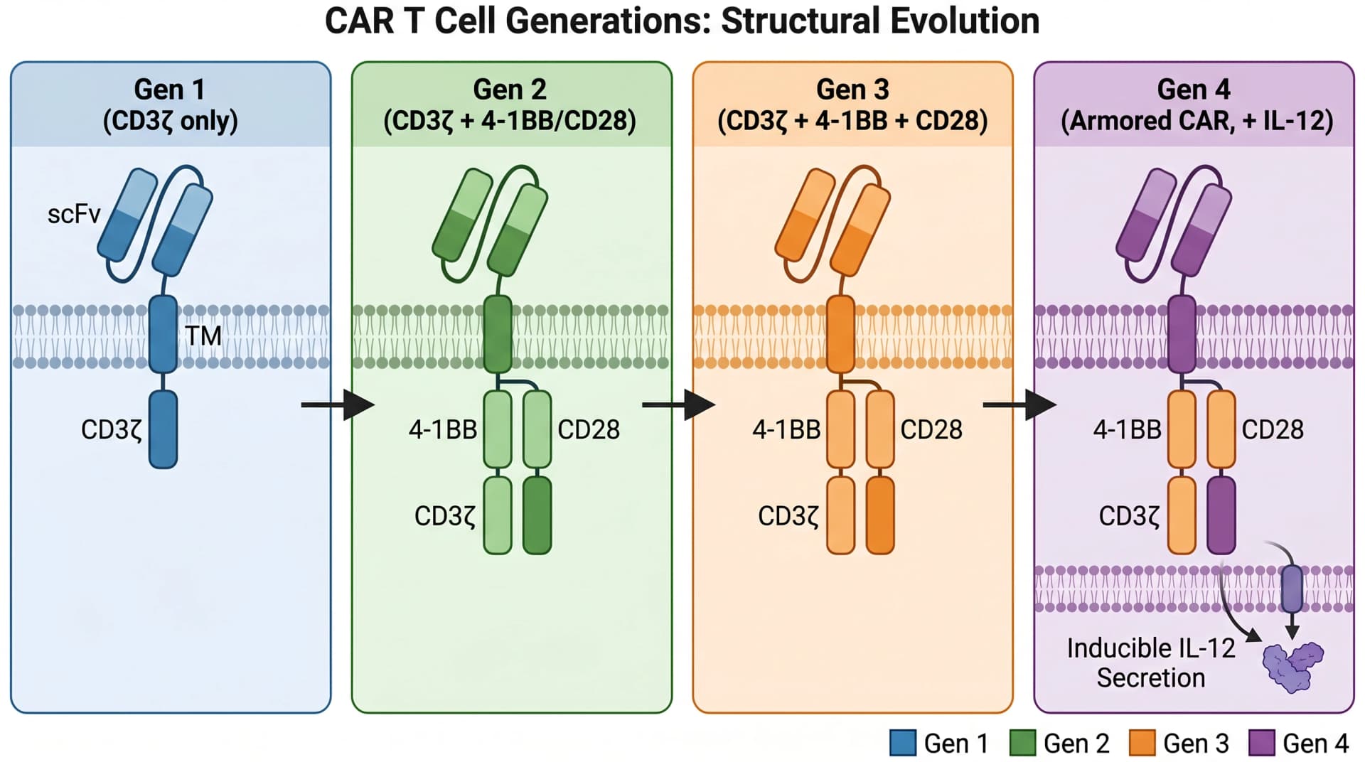
The visual challenge is making the differences obvious at a glance. Color-coding by generation works; arranging vertically with explicit labels for each new component works; using a "stack" metaphor where each generation adds a layer onto the previous works. What does not work is asking a generic image model to "draw 4 generations of CAR" without specifying which costimulatory domain is where — you will get a randomized result every time.
| Generation | Costimulatory Architecture | Clinical Status (2026) | Representative Products |
|---|---|---|---|
| Gen 1 | CD3ζ only — no costim | Largely abandoned (poor persistence) | None approved |
| Gen 2 | Single costim (4-1BB or CD28) + CD3ζ | Dominant clinical scaffold | Kymriah (4-1BB), Yescarta (CD28), Abecma, Carvykti, Breyanzi, Tecartus |
| Gen 3 | Dual costim (4-1BB + CD28) + CD3ζ | Clinical trials underway | None approved as of 2026 |
| Gen 4 ("armored") | Single/dual costim + CD3ζ + inducible cytokine (IL-12 or IL-18) | Early-phase trials | None approved as of 2026 |
Tip
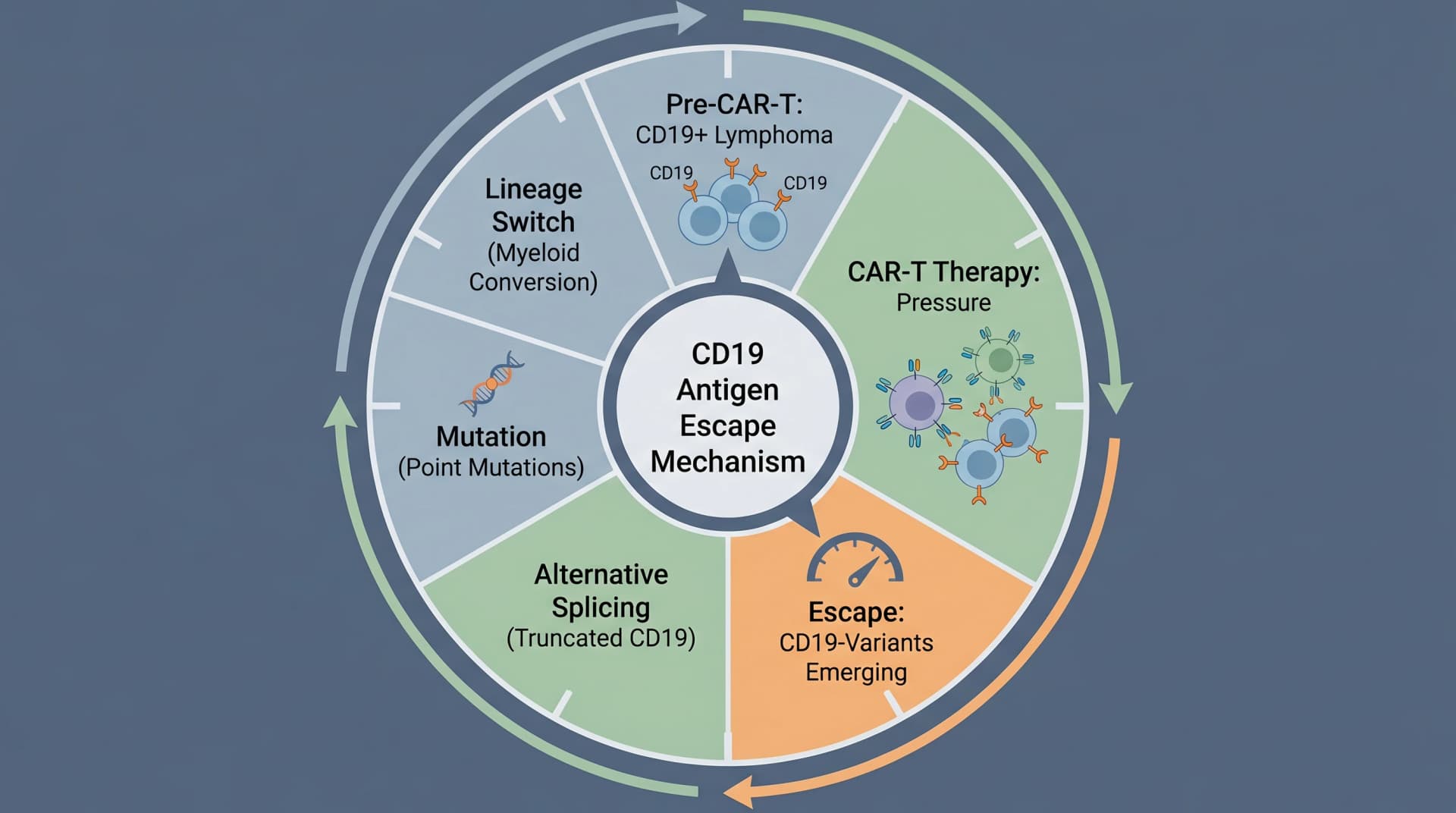
4. CD19, BCMA, and the Antigen Target Map: From DLBCL to Multiple Myeloma
CAR-T target selection has expanded dramatically beyond CD19. Each target maps to specific malignancies, and your poster needs to show the antigen on the tumor surface plus the rationale for choosing it.
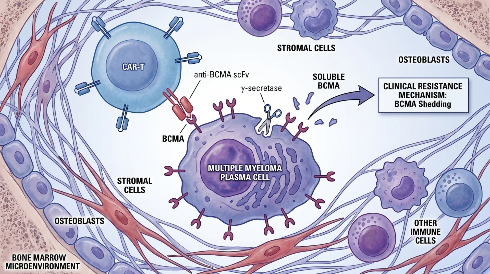
For multiple-myeloma-focused posters, the figure must show not just the BCMA antigen but the bone marrow microenvironment context — osteoblasts, stromal cells, and the γ-secretase shedding mechanism that produces soluble BCMA, a known clinical resistance pathway.
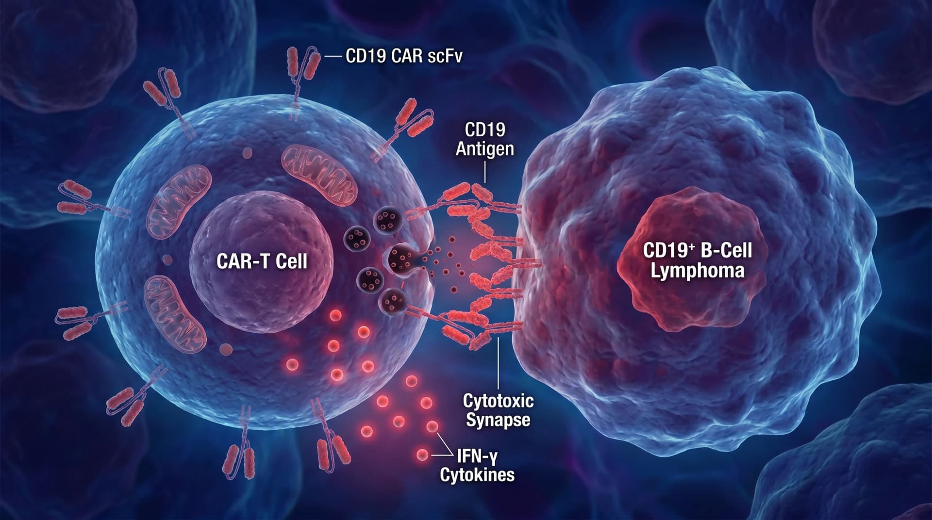
5. Bispecific Antibodies (BiTE) and the T-Cell Engager Family
Bispecific T-cell engagers are conceptually adjacent to CAR-T — both use redirected T-cell cytotoxicity — but the figure is fundamentally different because the redirecting molecule is a soluble antibody, not an engineered cell. EHA 2026 sees rapidly growing BiTE submissions across CD19 (blinatumomab), CD20 (mosunetuzumab, glofitamab), BCMA (teclistamab), and GPRC5D (talquetamab) targets.
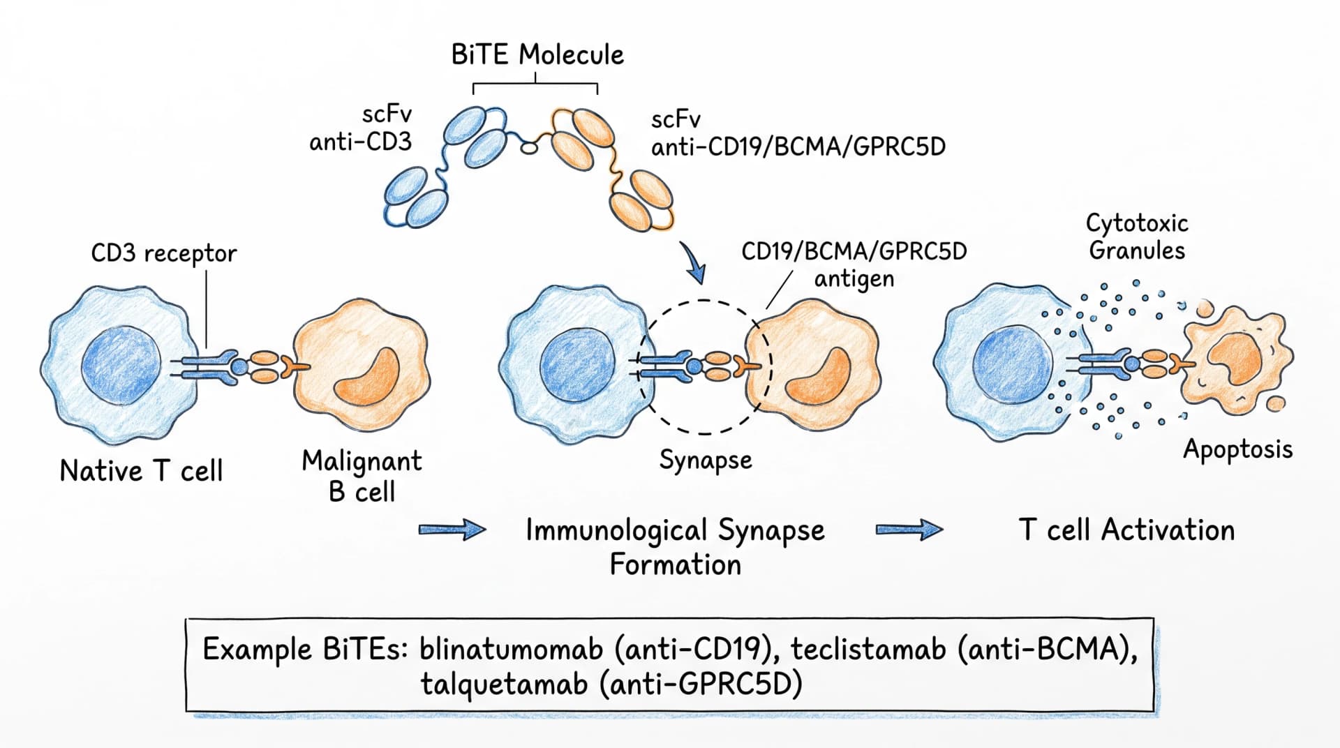
For a poster comparing CAR-T to BiTE in the same patient population (a common 2026 design), one panel shows the engineered cell construct, the other shows the bispecific antibody bridging — and a third panel can compare efficacy, persistence, and off-target safety side by side.
6. CAR-T Manufacturing Workflow Visualization
Reviewers want to see the manufacturing workflow because it tells them how reproducible your study is and where in the timeline your patients drop out. The canonical workflow has seven steps over 2–4 weeks.
- Leukapheresis — Patient T cells collected via apheresis
- T cell isolation — CD3+ enrichment, sometimes CD4/CD8 ratio adjustment
- Activation — CD3/CD28 bead stimulation
- Transduction — Lentiviral or retroviral delivery of the CAR gene
- Expansion — 7–10 day culture to clinically relevant numbers
- Cryopreservation and release testing — Quality control before product release
- Patient infusion — After lymphodepletion (typically fludarabine + cyclophosphamide)
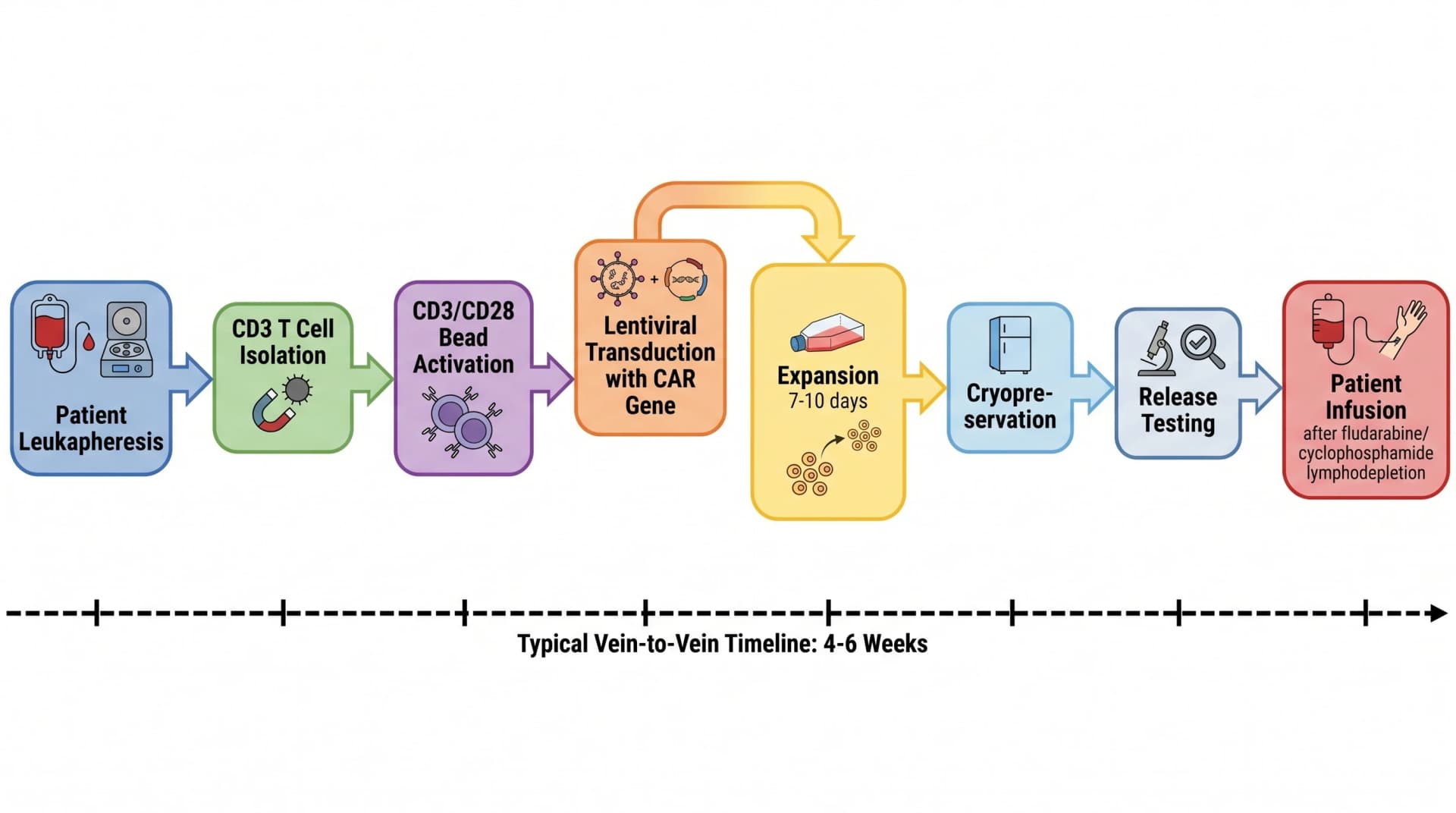
For autologous trials, the figure should include patient-specific timing markers — typical vein-to-vein time is 4–6 weeks, but bridging therapy windows shorten the actually-useful preparation time. Allogeneic ("off-the-shelf") products compress this dramatically and are worth a contrasting subpanel if your work addresses both.
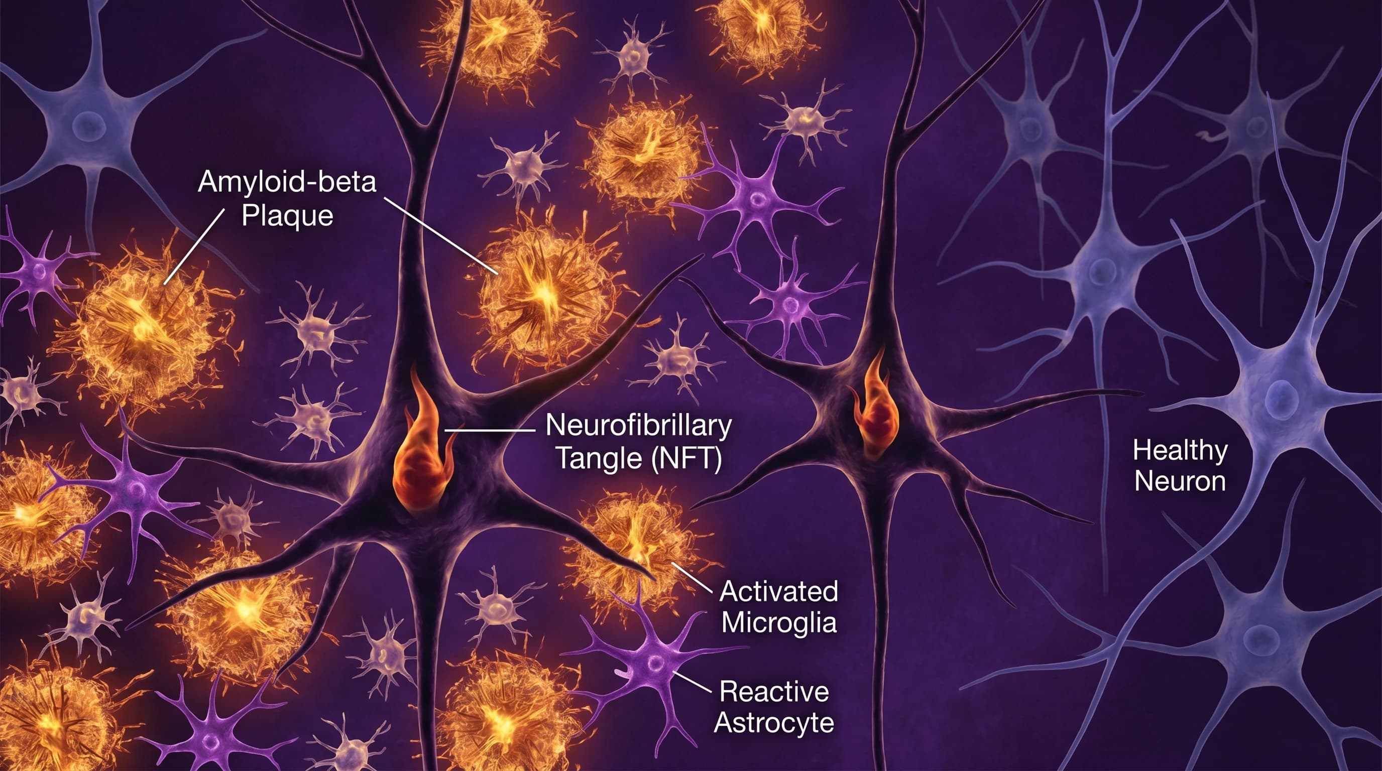
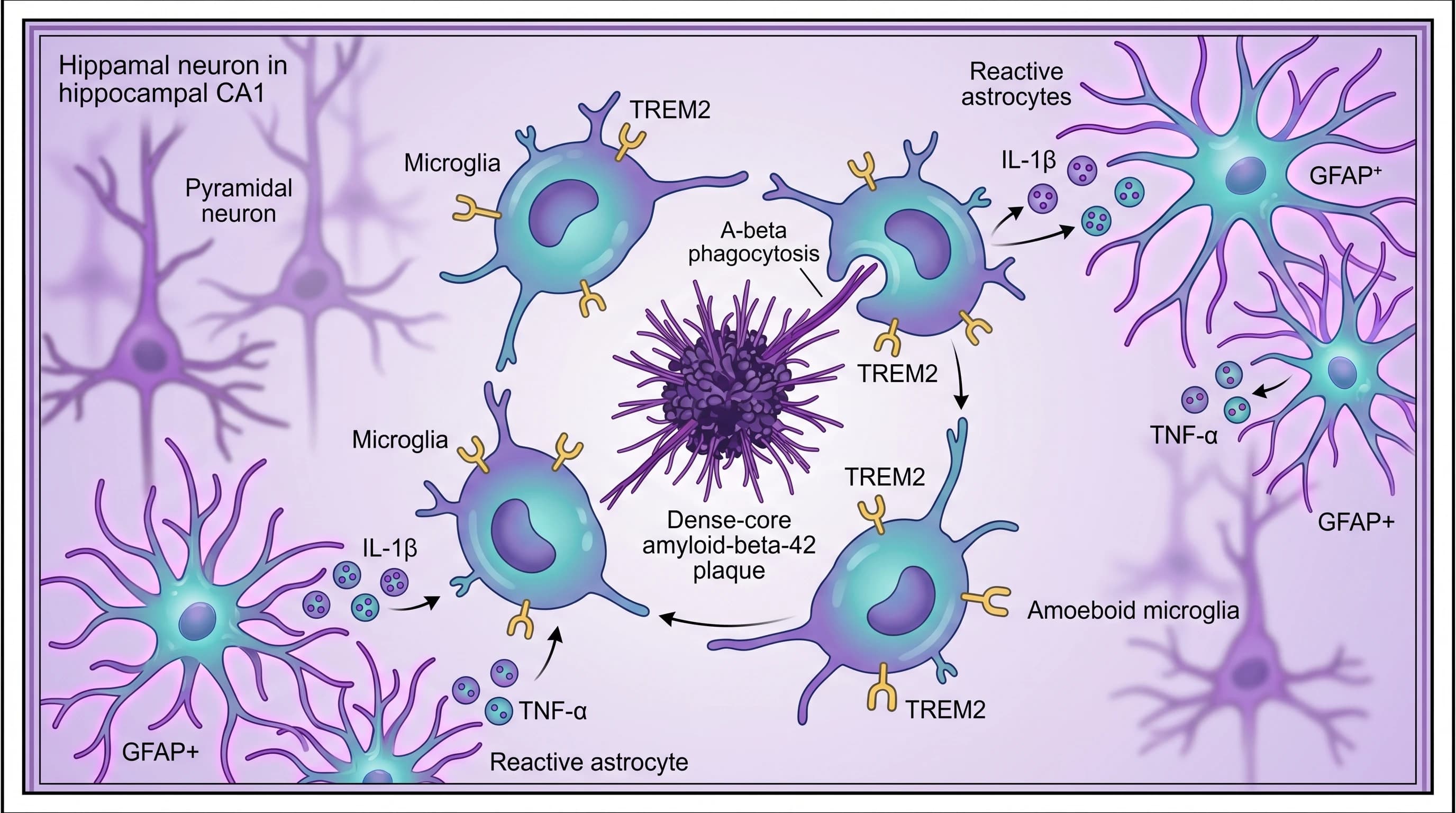
7. CRS and ICANS Pathophysiology Diagrams
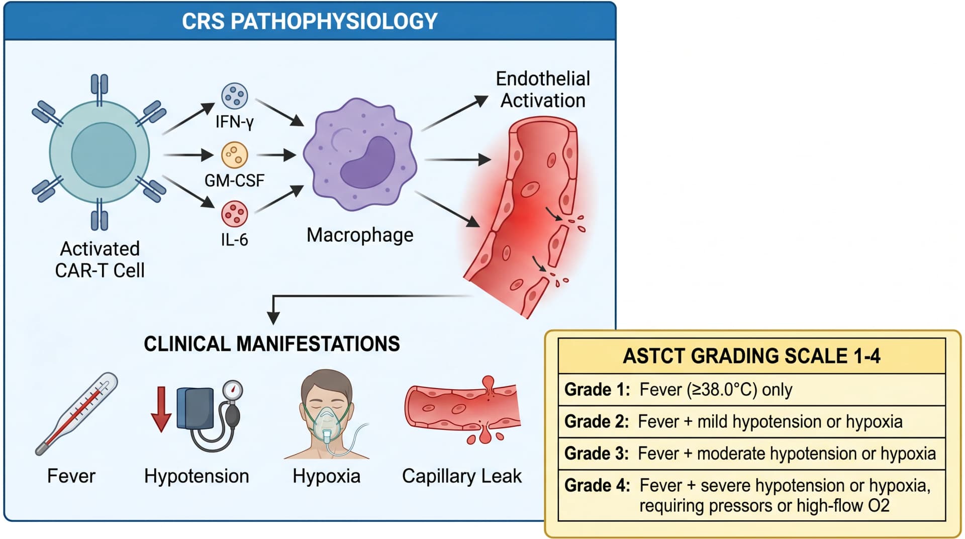
Cytokine release syndrome (CRS) and immune effector cell-associated neurotoxicity syndrome (ICANS) are the two signature toxicities of CAR-T therapy, and EHA reviewers expect every CAR-T poster to address them at least at the safety-data level. A pathophysiology figure converts a wall of cytokine names into a clear cascade.
ICANS pathophysiology is incompletely understood but appears to involve disruption of the blood-brain barrier and microglial activation; a figure for an ICANS-focused poster can show the BBB compromise schematically while explicitly noting that the upstream cytokine drivers are still being defined.
8. From Prompt to Publication-Ready: SciFig Workflow for CAR-T Mechanism Diagrams
Here is the part most CAR-T poster authors find genuinely transformative — and it is also where you find out why generic image models are not enough for this kind of figure.
If you have already tried drawing a Gen 2 CAR with GPT image or Midjourney, you have probably seen this: the model puts 4-1BB and CD28 in the wrong vertical order, or it gives you two ITAMs on the CD3ζ chain instead of three, or it labels the costimulatory domain "4-1BB" while drawing CD28's structure. You re-roll, and the next version makes a different mistake — maybe the scFv has three domains now, or the transmembrane segment floats above the membrane instead of crossing it. This is not a failure of one specific vendor; no generic image model today can reliably reach 100% accuracy on a CAR construct on the first try, because the model has not been built around the specific molecular grammar that hematologists use. And in CAR-T design, 99% accurate means 0% — an expert reviewer will spot a CD3ζ chain pointing the wrong way within seconds, and your entire mechanistic story collapses with that one error.
SciFig is built for this exact gap. Best-in-class image generation models bring the first-pass output to a high-fidelity starting point — the five-component CAR construct, the immunological synapse, the BiTE bridging — most of which is topologically correct on draft one. But for the precision details that matter most — domain ordering, ITAM motif count, costimulatory domain identity, transmembrane orientation — an editable vector canvas in the browser lets you click any label and rename it, drag any domain and reposition it, swap an entire costimulatory domain from 4-1BB to CD28 without rerolling the whole figure. The remaining precision gap closes in seconds, not minutes. And the entire workflow stays inside SciFig — one-click export to editable PPTX for your lab meeting, layered SVG for downstream editing, or 8K PNG for A0 poster printing without artifacting. There is no roundtrip to Illustrator, no figure reduction to a flat pixel image, no re-creation from scratch when a reviewer asks for one small change.
Here is the path. Copy this prompt verbatim into SciFig's Text-to-Figure tool to start your CAR construct figure:
Labeled diagram of CAR construct showing 5 components from outside
to inside: (1) anti-CD19 scFv antigen-binding domain with V_H and V_L
linked by flexible linker, (2) CD8α-derived hinge region, (3) single
alpha-helical transmembrane domain crossing phospholipid bilayer,
(4) intracellular 4-1BB costimulatory domain, (5) CD3ζ activation
signaling tail with 3 ITAM motifs. Clean vertical layout with callout
labels and leader lines, publication-ready style, blue/red color palette.
For the manufacturing workflow, the BiTE bridging, the CRS cascade, and the BCMA myeloma figures — copy the prompts in Section 10 below.
See AI Scientific Figure Generation in Action
Watch how researchers create publication-ready scientific figures from text descriptions.
Explore the Tool9. Common Mistakes When Drawing CAR-T Mechanism Diagrams
The errors that reviewers spot most often in CAR-T poster figures fall into five categories. Catch them before submission.
- Wrong number of scFv domains — Should be exactly two (V_H + V_L). One or three is the most common AI-generated error.
- Wrong number of CD3ζ ITAMs — Should be exactly three. Two or four is common.
- CD3ζ pointing extracellularly — The activation domain is intracellular by definition. Reversing it is a topology error.
- Conflating CAR with native TCR — A native T-cell receptor has α and β chains plus CD3 complex; a CAR is a single chimeric receptor. Drawing α/β chains on a CAR figure is a category error.
- Costimulatory domain mislabeled — 4-1BB structure shown with CD28 label or vice versa. Check that the structure matches the label.
10. Free Trial CTA + Related Reading: 5 Copy-Paste CAR-T Prompts
The five remaining SciFig prompts for the figures shown in this article. Copy any of them directly into Text-to-Figure:
Side-by-side comparison of 4 CAR generations from left to right.
Gen 1: CD3ζ only. Gen 2: CD3ζ + 4-1BB or CD28 costimulation. Gen 3:
dual costimulation (4-1BB + CD28). Gen 4 (armored CAR): adds inducible
IL-12 secretion. Each generation shown with full structural diagram,
horizontal panel layout, color-coded by generation, publication style.
CAR-T cell engineered with anti-BCMA scFv targeting BCMA-expressing
multiple myeloma plasma cell in bone marrow microenvironment. Show
surrounding stromal cells, osteoblasts, and other immune cells.
Highlight BCMA shedding via γ-secretase as a clinical resistance mechanism.
Schematic of bispecific T-cell engager (BiTE): two scFv arms, one
anti-CD3 binding native T cell, one anti-CD19 (or anti-BCMA for myeloma)
binding malignant B cell. Show formation of immunological synapse and
resulting T cell activation. Annotate with example drug names
(blinatumomab CD19, teclistamab BCMA, talquetamab GPRC5D).
Horizontal workflow diagram for autologous CAR-T manufacturing:
patient leukapheresis → CD3 T cell isolation → CD3/CD28 bead activation
→ lentiviral transduction with CAR gene → expansion 7-10 days →
cryopreservation → release testing → patient infusion after
fludarabine/cyclophosphamide lymphodepletion. Annotate typical
vein-to-vein timeline of 4-6 weeks.
Cytokine release syndrome (CRS) pathophysiology: activated CAR-T cell
releases IFN-γ, GM-CSF, and IL-6. Macrophages amplify the IL-6 cascade.
Show endothelial activation and resulting clinical manifestations:
fever, hypotension, hypoxia, capillary leak. Inset showing ASTCT
grading scale 1-4 with severity criteria.
Create Scientific Figures Now
Describe your scientific figure in natural language — get publication-ready illustrations in minutes.
Try Free